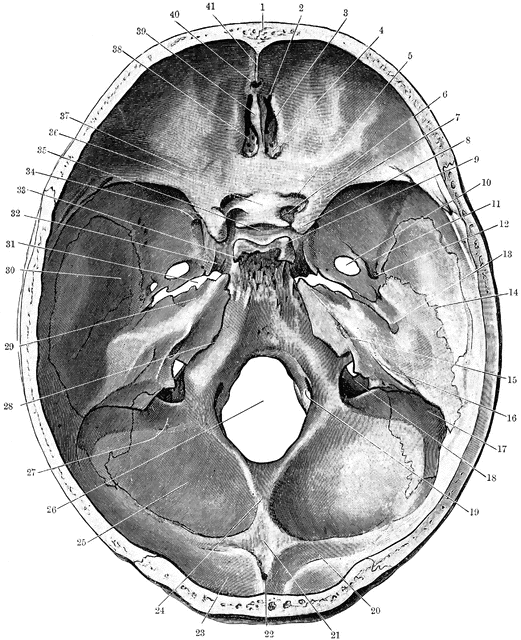
Base of the Skull Seen From Above
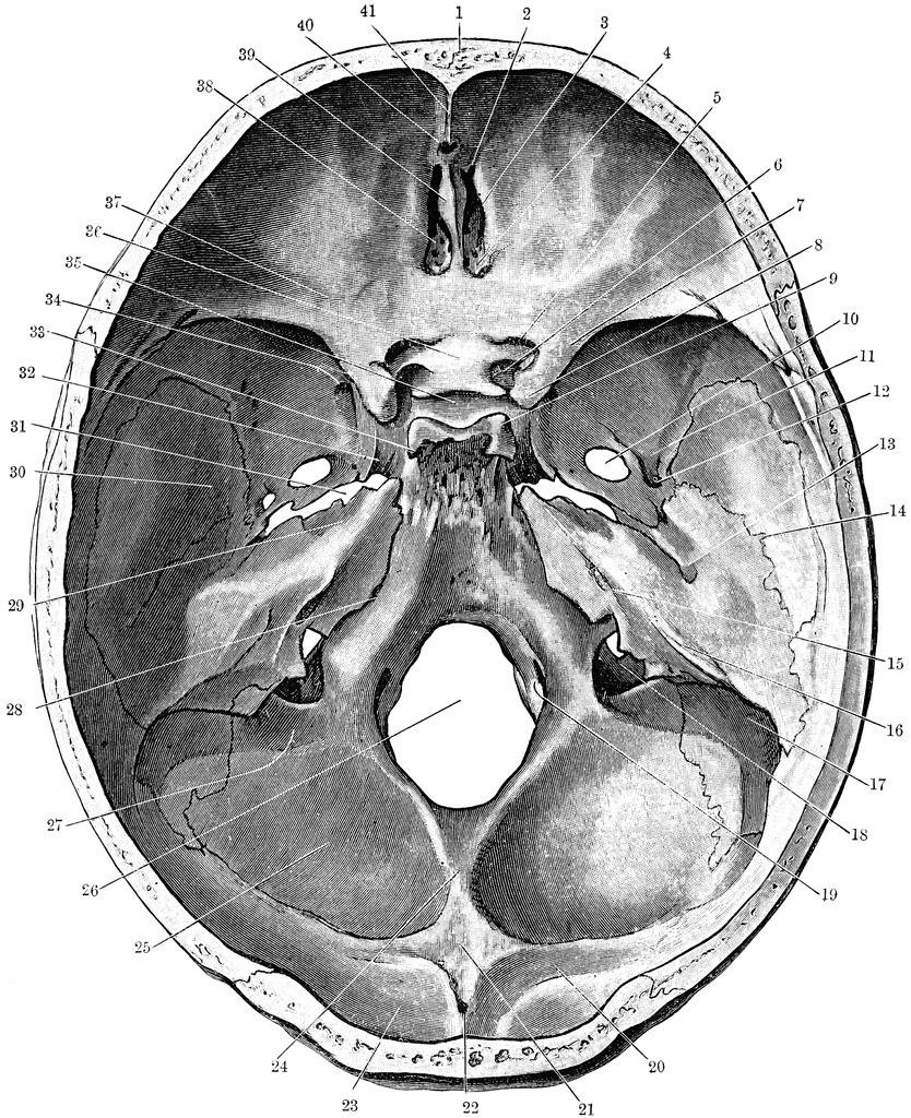
Shown is the base of the skull seen from above. Labels: 1, frontal bone; 2, slit for nasal nerve; 3, anterior ethmoidal foramen; 4, posterior ethmoidal foramen; 5, optic foramen; 6, foramen for internal carotid artery formed by anterior and middle clinoid processes; 7, lesser wing of sphenoid; 8, anterior clinoid process, in this case united on its inner side to the middle clinoid process; 9, posterior clinoid process; 10, foramen ovale; 11, groove for middle meningeal artery; 12, foramen spinosum; 13, Hiatus Fallopii; 14, line of petro-squamosal suture; 15, internal auditory meatus; 16, groove for superior petrosal sinus; 17, groove for sigmoid part of lateral sinus; 18, jugular foramen; 19, anterior condylic foramen; 20, groove for lateral sinus; 21, internal occipital protuberance; 22, ridge for attachment of falx cerebri; 23, fossa for the lodgment of the occipital lobe of the cerebrum; 24, ridge for attachment of the falx cerebelli; 25, fossa for the lodgment of the left cerebellar hemisphere; 26, foramen magnum; 27, groove for the sigmoid sinus turning into the jugular foramen; 28, groove for the inferior petrosal sinus running along the line of the suture between the petrous temporal and the basioccipital; 29, depression for the Gasserian ganglion; 30, middle cranial fossa for lodgment of temporal lobe of cerebrum; 31, foramen lacerum medium; 32, carotid groove; 33, dorsum sellae of sphenoid; 34, leads into foramen rotundum; 35, pituitary fossa; 36, olivary eminence of sphenoid; 37, anterior cranial fossa for lodgment of frontal lobes of cerebrum; 38, cribriform plate of ethmoid; 39, crista galli of ethmoid; 40, foramen caecum; 41, crest for attachment of falx cerebri.
Keywords
head, skull, cranium, "base of skull", "bones of the head", "facial bones", "bones of the face"Galleries
Human Skeletal SystemSource
Cunningham, D.J. Textbook of Anatomy (New York, NY: William Wood and Co., 1903)
Downloads
1957×2400, 3.4 MiB
834×1024, 397.8 KiB
521×640, 146.6 KiB
260×320, 37.8 KiB