Shown is the base of the skull seen from above. Labels: 1, frontal bone; 2, slit for nasal nerve; 3, anterior ethmoidal foramen; 4, posterior ethmoidal foramen; 5, optic foramen; 6, foramen for internal carotid artery formed by anterior and middle clinoid processes; 7, lesser wing of sphenoid; 8, anterior clinoid process, in this case united on its inner side to the middle clinoid process; 9, posterior clinoid process; 10, foramen ovale; 11, groove for middle meningeal artery; 12, foramen spinosum; 13, Hiatus Fallopii; 14, line of petro-squamosal suture; 15, internal auditory meatus; 16, groove for superior petrosal sinus; 17, groove for sigmoid part of lateral sinus; 18, jugular foramen; 19, anterior condylic foramen; 20, groove for lateral sinus; 21, internal occipital protuberance; 22, ridge for attachment of falx cerebri; 23, fossa for the lodgment of the occipital lobe of the cerebrum; 24, ridge for attachment of the falx cerebelli; 25, fossa for the lodgment of the left cerebellar hemisphere; 26, foramen magnum; 27, groove for the sigmoid sinus turning into the jugular foramen; 28, groove for the inferior petrosal sinus running along the line of the suture between the petrous temporal and the basioccipital; 29, depression for the Gasserian ganglion; 30, middle cranial fossa for lodgment of temporal lobe of cerebrum; 31, foramen lacerum medium; 32, carotid groove; 33, dorsum sellae of sphenoid; 34, leads into foramen rotundum; 35, pituitary fossa; 36, olivary eminence of sphenoid; 37, anterior cranial fossa for lodgment of frontal lobes of cerebrum; 38, cribriform plate of ethmoid; 39, crista galli of ethmoid; 40, foramen caecum; 41, crest for attachment of falx cerebri.

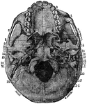
Base of the Skull Seen From Above

Shown is the base of the skull seen from above. Labels: 1, frontal bone; 2, slit for nasal nerve; 3,…
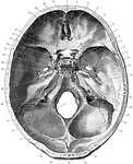
The base of the skull. "The lower jaw has been removed. At the lower part of the figure is the hard palate forming the roof of the mouth and surrounded by the upper set of teeth. Above this are the paired opening of the posterior nares, and a short way above the middle of the figure is the large median foramen magnum, with the bony convexities (or occipital condyles) which articular with the atlas, on its sides. It will be seen that the part of the skull behind the occipital condyles is about equal in size to that in front of them; in an ape the portion in front of the occipital condyles would be much larger than that behind them." —Newell, 1900

Base of the Skull

The base of the skull. "The lower jaw has been removed. At the lower part of the figure is the hard…

Foramina (openings) at the base of the skull.

Base of the Skull

Foramina (openings) at the base of the skull.