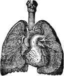
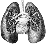
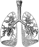
View of the bronchia and veins of the lungs, exposed by dissection, as well as the relative position of the lungs to the heart.

Bronchia and Veins of the Lungs

View of the bronchia and veins of the lungs, exposed by dissection, as well as the relative position…
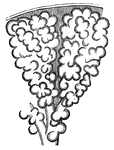
This diagram shows the bronchial tubes, with clusters of cells. The bronchioles are the first airway branches that no longer contain cartilage. They are branches of the bronchi, and are smaller than one millimetre in diameter.

Bronchioles

This diagram shows the bronchial tubes, with clusters of cells. The bronchioles are the first airway…
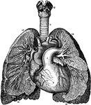
A view of the bronchia and blood vessels of the lungs, as shown by dissection, as well as the relative position of the lungs to the heart.

Relative Position of the Heart and Lungs

A view of the bronchia and blood vessels of the lungs, as shown by dissection, as well as the relative…

Front view of cartilages of larynx, trachea and Bronchi.

Front View of the Cartilages of the Larynx, Trachea and Bronchi.

Front view of cartilages of larynx, trachea and Bronchi.
"<em>A</em>, division of left bronchus to upper lobe; <em>B</em>, left branch of the pulmonary artery; <em>C</em>, left bronchus; <em>D</em>, left superior pulmonary vein; <em>E</em>, left inferior pulmonary vein; <em>F</em>, left auricle; <em>K</em>, inferior vena cava; <em>L</em>; division of the right bronchus to lower lobe; <em>M</em>, right inferior pulmonary vein; <em>N</em>, right superior pulmonary vein, <em>O</em>, right branch of the pulmonary artery; <em>P</em>, division of right bronchus to upper lobe; <em>R</em>, left ventricle; <em>S</em>, right ventricle." &mdash; Blaisedell, 1904

Lungs

"A, division of left bronchus to upper lobe; B, left branch of the pulmonary artery;…

The lungs and windpipe (trachea). Labels: 1, larynx; 2, windpipe (trachea); 3, right lung, showing bronchi and bronchial tube.

Lungs and Trachea

The lungs and windpipe (trachea). Labels: 1, larynx; 2, windpipe (trachea); 3, right lung, showing bronchi…

Mediastinal surfaces of the two lungs of a subject hardened by formalin injection. A, right lung. B, left lung. Labels: a, base; 2, fissure; 3, cardiac depression; 4, groove for innominate vein; 5, groove for innominate artery; 6, apex pulmonis; 7, groove for vena azygos major; 8, eparterial bronchus; 9, pulmonary artery (right); 10, fissure; 11, groove for aorta; 12, bronchus; 13, pulmonary artery (left); 14, apex pulmonis; 15, groove for left subclavian artery; 16, groove for left innominate vein; 17, cardiac depression; 18, fissure; 19, base.

Mediastinal Surfaces of the Lungs

Mediastinal surfaces of the two lungs of a subject hardened by formalin injection. A, right lung. B,…

Outline showing the general form of the larynx, trachea, and bronchi, as seen from behind. Labels: h, the great cornu of the hyoid bone; e,epiglottis; t, superior cornu of the thyroid cartilage ; t1, inferior cornu of the thyroid cartilage; a, points to the back of both the arytenoid cartilages, which are surmounted by the cornicula; c, the middle ridge on the back of the cricoid cartilage; tr, the posterior membranous part of the trachea; b, b1, right and left bronchi.

Back View of Respiratory Apparatus

Outline showing the general form of the larynx, trachea, and bronchi, as seen from behind. Labels: h,…

Outline showing the general form of the larynx, trachea, and bronchi, as seen from front. Labels: h, the great cornu of the hyoid bone; e,epiglottis; t, superior and inferior cornu of the thyroid cartilage; c, middle of the cricoid cartilage; tr, the trachea, showing sixteen cartilaginous rings; b, the right bronchus; b1, the left bronchus.

Front View of Respiratory Apparatus

Outline showing the general form of the larynx, trachea, and bronchi, as seen from front. Labels: h,…

"The respiratory mechanism consists of the lungs, a series of minute air chambers with a network of capillaries in the wall, the air passages from the air chambers of the lngs to the outer air, and the chest walls with their muscles, which act like bellows and change the ai r in the lungs. Let us begin with the air passages. There are first the nose and mouth; these join the upper part of the gullet, known as the pharynx. From the pharnyx arises the windpipe (trachea); this passes through the voice box (larynx) into the chest cavity; there it divides into two passages (the bronchi); the bronchi go on dividing again and again, generally into two; the ultimate divisions (the bronchioles) open into clusters of air chambers."&mdash;(Charles Leonard-Stuart, 1911)

Respiratory Mechanism

"The respiratory mechanism consists of the lungs, a series of minute air chambers with a network of…
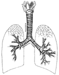
"Larynx, trachea, and bronchi, showing the manner of division, and the rings of cartilage." &mdash; Tracy, 1888

Respiratory system

"Larynx, trachea, and bronchi, showing the manner of division, and the rings of cartilage." —…

"Larynx, Trachea, and the Bronchi. (Front view.) <em>A</em>, epiglottis; <em>B</em>, thyroid cartilage; <em>C</em>, cricothyroid membrane, connecting with the cricoid cartilage below, all forming the larynx; <em>D</em>, rings of the trachea." &mdash; Blaisedell, 1904

Trachea

"Larynx, Trachea, and the Bronchi. (Front view.) A, epiglottis; B, thyroid cartilage;…

The trachea and bronchi. The thyroid body is indicated by a dotted line.

Trachea and Bronchi

The trachea and bronchi. The thyroid body is indicated by a dotted line.

"Bifurcation of trachea, and bronchi, viewed from below; a, pessulus, the bolt-bar, or "bone of divarication"; b, b, next succeeding tracheal half-rings.Elliot Coues, 1884

The Trachea of a Rook

"Bifurcation of trachea, and bronchi, viewed from below; a, pessulus, the bolt-bar, or "bone of divarication";…