Circulation in frog's foot under a microscope. Labels: A, walls of capillaries; B, tissue of web lying between the capillaries; C, cells of epidermis covering web (these are only shown in the right-hand and lower part of the field; in the other parts of the field the focus of the microscope lies below the epidermis); D, nuclei of these epidermic cells; E, pigment cells contracted, not partially expanded; F, red blood corpuscle (oval in the frog) passing along capillary -- nucleus not visible; G, another corpuscle squeezing its way through a capillary, the canal of which is smaller than its own transverse diameter; H, another bending as it slides round a corner; K, corpuscle in capillary seen through the epidermis; I, white blood corpuscle.

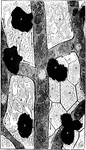
Circulation in a Frog's Foot

Circulation in frog's foot under a microscope. Labels: A, walls of capillaries; B, tissue of web lying…