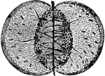
"Aldrovanda vesiculosa. Leaf pressed open and enlarged, showing glands, sensitive filaments, and quadrifid hairs." — The Encyclopedia Britannica, 1893

Aldrovanda

"Aldrovanda vesiculosa. Leaf pressed open and enlarged, showing glands, sensitive filaments, and quadrifid…

"Abdominal Plate (worker of Apis), under side, third segment. W, wax-yielding surface, covering true gland; s, septem, or carina; wh, webbed hairs." — The Encyclopedia Britannica, 1910

Bee Abdomen

"Abdominal Plate (worker of Apis), under side, third segment. W, wax-yielding surface, covering true…
A diagram of the digestive system.

Digestive System

A diagram of the digestive system.

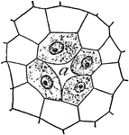
"Young resin gland of fir: <em>a</em>, duct, an intercellular space formed by the separation of the four nucleate cells." &mdash; Coulter, 1910

Resin gland of a fir

"Young resin gland of fir: a, duct, an intercellular space formed by the separation of the…

"The inner coat of the stomach has its surface honeycombed with millions of little pits. We have all seen this in tripe. In the floor of each of these tiny pits a number of tubes open. These are the openings of the <em>gastric glands</em>." &mdash; Blaisedell, 1904

Gastric gland

"The inner coat of the stomach has its surface honeycombed with millions of little pits. We have all…

Gland(g) from the upper surface of the leaf of lilac: e, epidermis, c, cuticle, p, palisade cells.

Gland

Gland(g) from the upper surface of the leaf of lilac: e, epidermis, c, cuticle, p, palisade cells.

"Diagram to show the working parts of a gland. v and a are blood tubes with thin-walled branches around the parts of the gland c. These take material from the blood and, after changing it, send it to the mouth, stomach, or other places before the duct i." &mdash;Davison, 1910

Gland

"Diagram to show the working parts of a gland. v and a are blood tubes with thin-walled branches around…
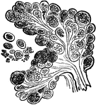
A racemose gland, which is a gland where the ducts are branched and clustered like grapes.

Racemose Gland

A racemose gland, which is a gland where the ducts are branched and clustered like grapes.
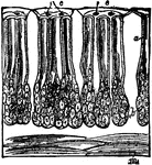
A simple secreting gland. Secreting glands serve to secrete (i.e., separate out some substance from the blood). They vary in size and structure.

Simple Gland

A simple secreting gland. Secreting glands serve to secrete (i.e., separate out some substance from…

"The convoluted gland is seen surrounded by fat cells and may be traced through the true skin to its outlet in the horny layers of the outer skin." &mdash; Blaisedell, 1904

Sweat gland

"The convoluted gland is seen surrounded by fat cells and may be traced through the true skin to its…

Diagram showing development of glands: A, a mere dimple in the surface; B, enlargement by division; C, enlargement by dilation; D, a combination of B and C; E, a racemose gland; F, development of method of E; G, a single tube intricately coiled.

Development of Glands

Diagram showing development of glands: A, a mere dimple in the surface; B, enlargement by division;…
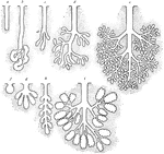
Diagram showing types of glands. a-e, tubular; f-i, alveolar or saccular; a, simple; b, coiled; c-d, increasingly complex compound tubular; e, tuboaiveolar; f, simple; g-h, progressively complex compound alveolar.

Types of Glands

Diagram showing types of glands. a-e, tubular; f-i, alveolar or saccular; a, simple; b, coiled; c-d,…

"<em>A</em>, a fold of peritoneum; <em>B</em>, lacteals and lymphatic glands; <em>C</em>, veins of intestines; <em>R.C.,</em>, receptacle of the chyle (receptaculum chyli); <em>P.V.</em>, portal vein; <em>H.V.</em>, hepatic veins; <em>S.V.C.</em>, superior vena cava; <em>R.A.</em>, right auricle of the heart; <em>I.V.C.</em>, inferior vena cava." &mdash; Blaisedell, 1904

Intestinal absorption

"A, a fold of peritoneum; B, lacteals and lymphatic glands; C, veins of intestines;…
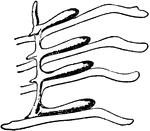
"The right coxal gland of Limulus polyphemus, Latr. a2 to a5, Posterior borders of the chitinous bases of the coxae of the second, third, fourth and fifth prosomatic limbs. b, Longitudinal lobe or stolon of the coxal gland. c, It's four transverse lobes or outgrowths corresponding to the four coxae." &mdash; The Encyclopedia Britannica, 1910

Limulus Polyphemus

"The right coxal gland of Limulus polyphemus, Latr. a2 to a5, Posterior borders of the chitinous bases…

"Consisting of a very fine network of fibrils, around which are cells of various sizes." — Blaisedell, 1904

Connective Tissue from a Lymphatic Gland

"Consisting of a very fine network of fibrils, around which are cells of various sizes." — Blaisedell,…
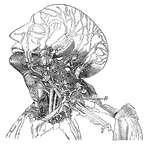
This diagram shows the lumphatics of the head and neck. It shows the glands, and B, the thoracic duct as it empties into the left innominate vein at the junction of the left jugular and subclavian veins.

Lymphatics

This diagram shows the lumphatics of the head and neck. It shows the glands, and B, the thoracic duct…
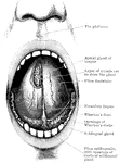
Open mouth with tongue raised, and the sublingual and apical glands exposed.

Mouth and Tongue

Open mouth with tongue raised, and the sublingual and apical glands exposed.

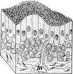
"A tiny block of tissue from the membrane lining the inner surface of the nose. Note the hundreds of cillia and the gland m which gives out the slippery white or colorless fluid called mucus." —Davison, 1910

Section of Nasal Tissue

"A tiny block of tissue from the membrane lining the inner surface of the nose. Note the hundreds of…
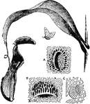
"Pitcher of Nepenthes distillatoria. A, honey-gland from attractive surface of lid; B, digestive gland from interior of pitcher, in pocket-like depression of epidermis, opening downwards; C, transverse section of the same. A, B, and C magnified about 100 diameters." &mdash; The Encyclopedia Britannica, 1893

Nepenthes

"Pitcher of Nepenthes distillatoria. A, honey-gland from attractive surface of lid; B, digestive gland…

"Vertical section of part of the rind of the Orange, showing glands containing volatile oil, R, R, R, surrounded by cells." &mdash; Encyclopedia Britanica, 1893

Orange Rind

"Vertical section of part of the rind of the Orange, showing glands containing volatile oil, R, R, R,…

"Surface of Palm of the Hand, showing Openings of Sweat Glands and Grooves between Papillae of the Skin. In the smaller figure the same epidermal surface is shown as seen with the naked eye." &mdash; Blaisedell, 1904

Surface of the palm

"Surface of Palm of the Hand, showing Openings of Sweat Glands and Grooves between Papillae of the Skin.…
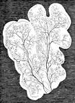
In mammalia, each salivary gland first appears as a simple canal with bud-like processes, lying in a gelatinous nidus or blastema, and communicating with the cavity of the mouth. As the development of the gland advances, the canal becomes more and more ramified, increasing at the expense of the blastema in which it is still enclosed. the branches or salivary ducts constitute an independent system of closed tubes. Shown are lobules of the parotid, with salivary ducts, in the embryo of the sheep at a more advanced stage.

Lobules of Parotid of a Sheep

In mammalia, each salivary gland first appears as a simple canal with bud-like processes, lying in a…
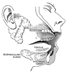
"While the food is being chewed it is moistened by saliva, or spittle, which flows into the mouth from six little glands. They are known as salivary glands.  There are three of them on each side." &mdash; Blaisedell, 1904

Salivary glands

"While the food is being chewed it is moistened by saliva, or spittle, which flows into the mouth from…
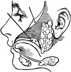
"There are three pairs of salivary glands. One pair lies under the tongue; one pair is found under the corners of the lower jaw; and the other pair is found in front of and below the ears." &mdash; Ritchie, 1918

Salivary glands

"There are three pairs of salivary glands. One pair lies under the tongue; one pair is found under the…
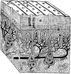
"<em>A</em>, simple tubular gland; <em>B</em>, gland with mouth shut and sac formed; <em>C</em> gland with a coiled tube; <em>D</em>, plan of part of a racemose gland." &mdash; Blaisedell, 1904

Secreting glands

"A, simple tubular gland; B, gland with mouth shut and sac formed; C gland…

"<em>A</em>, outer layer of cuticle; <em>B</em>, deeper layer of cuticle; <em>C</em>, duct of sweat gland; <em>D</em>, true skin; <em>E</em>, sublayer of true skin, with columnar cells. The blood vessels are injected to show black." — Blaisedell, 1904

Cross-Section of the Skin

"A, outer layer of cuticle; B, deeper layer of cuticle; C, duct of sweat…

"A block out of the skin. a, dead part and d live part of the epidermis; e, sweat glands; n, nerve endings." &mdash;Davison, 1910

Section of skin

"A block out of the skin. a, dead part and d live part of the epidermis; e, sweat glands; n, nerve endings."…
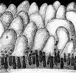
"<em>A, B,</em> glands seen in vertical section with their orifices at <em>C</em> opening upon the membrane between the villi; <em>D</em>, villus." &mdash; Blaisedell, 1904

Glands and villi of the small intestine

"A, B, glands seen in vertical section with their orifices at C opening upon the membrane…

"Anatomy of the Snail: a, the mouth; bb, foot; c, anus; dd, lung; e, stomach, covered above by the salivary glands; ff, intestine; g, liver; h, heart; i, aorta; j, gastric artery; l, hepatic artery; k, artery of the foot; mm, abdominal cavity, supplying the place of a cenous sinus; nn, irregular canal in communication with the abdominal cavaty, and carrying the blood to the lung; oo, vessel carrying the blood from the lung to the heart." &mdash; Chambers, 1881

Snail Anatomy

"Anatomy of the Snail: a, the mouth; bb, foot; c, anus; dd, lung; e, stomach, covered above by the salivary…
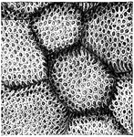
"The Inner Surface of the Stomach, from which the the Epithelium has been removed, showing the Openings of Gastric Glands." &mdash; Blaisedell, 1904

Inner surface of the stomach

"The Inner Surface of the Stomach, from which the the Epithelium has been removed, showing the Openings…
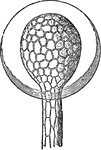
"Glands of Sundew magnified. External aspect with drop of secretion." &mdash; The Encyclopedia Britannica, 1893

Sundew

"Glands of Sundew magnified. External aspect with drop of secretion." — The Encyclopedia Britannica,…

"Glands of Sundew magnified. Internal structure." &mdash; The Encyclopedia Britannica, 1893

Sundew

"Glands of Sundew magnified. Internal structure." — The Encyclopedia Britannica, 1893

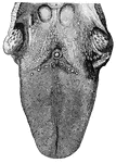
<em>A</em>, epiglottis; <em>B</em>, glands at the base of tongue; <em>C</em>, tonsil; The various kinds of papillae are plainly shown." &mdash; Blaisedell, 1904

Tongue

A, epiglottis; B, glands at the base of tongue; C, tonsil; The various kinds…

"Sensitive glands of Dionaea muscipula, x 300." &mdash; The Encyclopedia Britannica, 1893

Venus Fly-trap

"Sensitive glands of Dionaea muscipula, x 300." — The Encyclopedia Britannica, 1893

Section of the head of a serpent. a, poison fangs; b, poison glands; c, conductor for the poison; d, muscles of the jaws; e, tendon of the muscles; s, salivary glands.

Cross section of a viper head

Section of the head of a serpent. a, poison fangs; b, poison glands; c, conductor for the poison; d,…