"Portions of the rotting pulp were placed on a microscopic slide, divided into hundredths and thousandths of an inch. Fig. 21, A B, represents such a scale, the larger division, A B, representing the one-hundredth of an inch, and the smaller subdivision the one-thousandth of an inch; 7, 8, 9, 10, 11, 12, represent the cells of which the apples are mostly composed. The granular dottings represent apple starch. The branching cellular structure represents the mycelium of a fungus penetrating the cells. At 9, three small starch granuals are represented in a line, and are confined within the division of the one-thousandth of an inch." -Watts, 1874

Microscopic view of a fermented apple

"Portions of the rotting pulp were placed on a microscopic slide, divided into hundredths and thousandths…
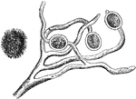
Illustration of a fungus named Atrotogus hydnosporus. "Considered by Berkeley and others to be probably a secondary form of fruit (spores) of the potato-fungus itself." -Watts, 1874

Fungus

Illustration of a fungus named Atrotogus hydnosporus. "Considered by Berkeley and others to be probably…

This illustration "represents its mycelium growth; 2,2 its budding cells, which terminate in fruit cells; 3, 3, 3, 3, bearing spores which germinate; 4, 4, are fungoid aggregations, which throw out filaments; their relation to the black mycelium is not represented thus far; 5 represents what appears to be a cross section of the asci or fruit, when perfectly formed and cut through its greatest diameter crosswise." -Watts, 1874

Stages of fungus growth

This illustration "represents its mycelium growth; 2,2 its budding cells, which terminate in fruit cells;…
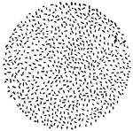
"Showing the Effect of Variations in Temperature on Bacteria Growth. a, a single bacterium; b, its progeny in twenty-four hours at fifty degrees; c, its progeny in twenty-four hours at seventy degrees." — Blaisedell, 1904

Bacteria Growth

"Showing the Effect of Variations in Temperature on Bacteria Growth. a, a single bacterium; b, its progeny…
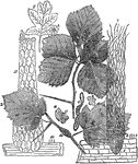
"Under the power of about 90 diameters the general character of the peridia is seen. They are densely aggregated, elongated, submerged, pale-brown, irregularly torn. The sporidia are copius...Nos. 1 and 2, indicate the general points of growth of this fungus. I find frequently on the leaf-ribs and terminal points of the leaves, and very often dispersed over the smooth parts of the leaf; sometimes, although rarely, the peridia are on the upper surface which, when matured, resembles net work. At the juncture of the leaf (see 4) the cells of the peridia are nearly round, at 5, oblong. From 3 to 4 the cellular structure is of light Vandyke brown; at 5, a pale yellow...6 represents the appearance of the peridia as see by the naked eye; 7, their general arrangements and their groupings on the leaves; 8, three cells, showing the parts of which the peridia are composed being magnified about 125 diameters." -Watts, 1874

Cellular structure of peridia

"Under the power of about 90 diameters the general character of the peridia is seen. They are densely…
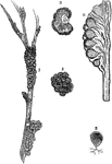
Illustrations depicting a black knot of a plum. "1...represents the general appearance of the black-knot of the plum; 2, a cross-section; 2, an enlarged view of it, showing indentations on the external surface of the conceptacles or perithecia of the fungus; 4, a longitudinal section of the black-knot and branch of a plumtree...5, a typical representation of the perithecia." -Watts, 1874

Black knot of the plum

Illustrations depicting a black knot of a plum. "1...represents the general appearance of the black-knot…

"In answer to a communication of mine, Professor C. H. Peck, botanist, of Albany new York, informs me that he has found sacks filled with spores with in the perithecia of black-knot, and has furnished me with a sketch of the sack as seen by him, (see U, Fig. 17.) I represents a highly magnified view of the true spore of this for of Shaeria." -Watts, 1874

Black knot of the plum

"In answer to a communication of mine, Professor C. H. Peck, botanist, of Albany new York, informs me…
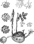
"It is not unusual to fine a decayed spot in the center of potatoes otherwise apparently in good condition. A microscopic examination a portion of the diseased part will show that the decay commenced where the vascular bundles concentrate. At that point the air is in greater volume than elsewhere. Although these mineral acids and caustic alkalies dissolve starch granuals, they do not affect the cellulose cells which contain the starch. When such spots are exposed to the atmosphere the fungus blue-mold forms on the surface. This disease, therefore, has not relation to potato-rot, as ordinarily understood. f represents the mycelium (roots) of blue-mold, Penicillium glaucum, and G that of potato-rot, Peronospora infestans." -Watts, 1874

Potato Diseases at a Microscopic Level

"It is not unusual to fine a decayed spot in the center of potatoes otherwise apparently in good condition.…

Illustration of a potato infested with Peronospora infestans also known as 'potato-rot' observed from a microscopic level.

Potato rot

Illustration of a potato infested with Peronospora infestans also known as 'potato-rot' observed from…
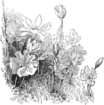
A perennial, herbaceous flowering plant native to eastern North America from Nova Scotia, Canada southward to Florida, United States.

Flowering Sanguinaria

A perennial, herbaceous flowering plant native to eastern North America from Nova Scotia, Canada southward…

A growth stage of a wild oats seedling, showing later growth.

Wild Oats Seedlng

A growth stage of a wild oats seedling, showing later growth.