Clipart tagged: ‘Human eye’
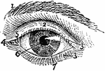
Exterior of Left Human Eye
"Exterior of Left Human Eye. 1, supercilium, or eyebrow; 2, palpebra superior, or upper eyelid; 3, 3,…
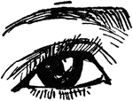
Human Eye
"Diagrammatic horizontal section of the eye of man. c, cornea; ch. choroid (dotted); C. P, ciliary processes;…
Median Vertical Anteroposterior Section of Eye
"Human Eye, in Median Vertical Anteroposterior Section. (Ciliary processes shown, through not all lying…
Eyeball
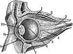
Section through the closed left eye. 1. Lifting muscle 2. Upper Straight Muscle 3. Optic Nerve 4. Fatty…
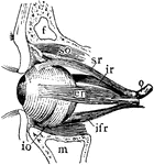
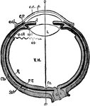
Muscles of Left Eyeball
"Muscles of Left Human Eyeball. so, superior oblique, passing through a trochlea or pulley; io, inferior…
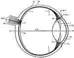
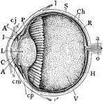
Human Eye
This diagram shows a side view of the right eye of man. a.c., central artery; a.h., aqueous humor; b.,…