Clipart tagged: ‘lachrymal’
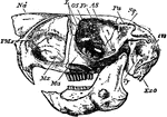
Cape Jumping Hare
"Side view of skull of Cape Jumping Hare. Pmx, premaxilla; Mx, maxilla; Ma, malar; Fr, frontal; L, lachrymal;…
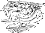
Cod Skull
"The skull of a cod. b, branchiostegal rays born on c.h., the ceratohyal bone; d, dentary portion of…
Eye and Lachrymal Gland
Front view of left eye, with eyelid partly removed to show lachrymal gland (tear-producing gland), and…
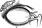
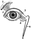
Eyeball
"The Relative Position of the Lachrymal Apparatus, the Eyeball, and the Eyelids. A, lachrymal canals,…
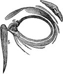
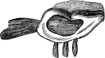
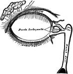
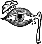
Eyelids, Viewed from the Front
The eyelids viewed from before; a,a, the lachrymal canals; b, the lachrymal sack. The lachrymal sac…
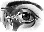
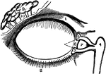
Lachrymal Apparatus
The lachrymal apparatus (the skin of the lids has been removed), which functions in tear production.


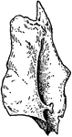
Inner Aspect of Lachrymal Bone
Right lachrymal bone, inner aspect. Upper part completes anterior ethmoidal cells, lower looks into…
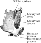
Human Lachrymal Facial Bone
Lachrymal Bone. The lachrymal are the smallest and most fragile bones fo the face. They are situated…
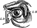
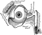
View of the Lachrymal Gland and Nasal Duct
View of the lachrymal gland and nasal duct. Labels: 1, The lachrymal gland. 2, Ducts leading from the…
The Lacrimal Gland
The lacrimal (lachrymal) gland is an oval gland in the orbital portion of the frontal bone. Its tear…
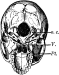
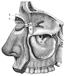
Base of the Skull
The base of the skull. The lower jaw has been removed. At the lower part of the figure is the hard palate…
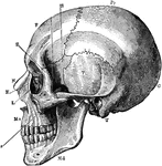
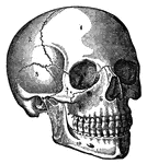
Side View of the Skull
A side view of the skull. Labels: O, occipital bone; T, temporal; Pr, parietal; F, frontal; S, sphenoid;…