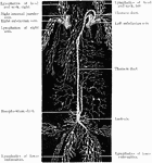
"<em>A</em>, a fold of peritoneum; <em>B</em>, lacteals and lymphatic glands; <em>C</em>, veins of intestines; <em>R.C.,</em>, receptacle of the chyle (receptaculum chyli); <em>P.V.</em>, portal vein; <em>H.V.</em>, hepatic veins; <em>S.V.C.</em>, superior vena cava; <em>R.A.</em>, right auricle of the heart; <em>I.V.C.</em>, inferior vena cava." &mdash; Blaisedell, 1904

Intestinal absorption

"A, a fold of peritoneum; B, lacteals and lymphatic glands; C, veins of intestines;…
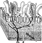
"A tiny block cut from the wall of the intestine showing villi and the mouths of glands at a; b, villus cut open to show the lacteal e and blood tubes m for absorbing food." —Davison, 1910

Section of Intestine wall

"A tiny block cut from the wall of the intestine showing villi and the mouths of glands at a; b, villus…

"The lymph vessels of the body. rc, the thoracic duct; lac, the lacteals taking the lymph and fatty part of food from the intestines." —Davison, 1910

Lymph Vessels

"The lymph vessels of the body. rc, the thoracic duct; lac, the lacteals taking the lymph and fatty…
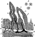
Diagram of the principle groups of the Lymphatic vessels.

Lymphatic Vessels

Diagram of the principle groups of the Lymphatic vessels.

Vertical section of the intestinal mucous membrane of the rabbit. Two villi are represented, in one of which the dilated lacteal alone is shown, in the other the blood vessels and lacteal are both seen injected, the lacteal white, the blood vessels dark; a, the lacteal vessels of the villi; a', horizontal lacteal, which they join; b, capillary blood vessels in one of the villi; c, small artery; d, vein; e, the epithelium covering the villi; g, tubular glands or crypts of Lieberkuhn, some divided down the middle, others cut more irregularly; i, the submucous layer. A, cross-section of three tubular glands more highly magnified.

Rabbit's Intestinal Mucous Membrane

Vertical section of the intestinal mucous membrane of the rabbit. Two villi are represented, in one…

"In the figure on the left are seen the artery and vein of a villus. In the right figure are represented the central lacteal of the villus, with the plexus of lymph vessels at the base. The mucous, submucuous, muscular, and serous coats are indicated at the base of the right-hand figure.  The epithelium of each villus is well shown." &mdash; Blaisedell, 1904

Transverse section of the small intestine

"In the figure on the left are seen the artery and vein of a villus. In the right figure are represented…

"The lacteals conduct the chyle from the intestines into numerous glands nearby, called the mesenteries, from which a fewer number of lacteals lead the chyle into one large tube in front of the spinal column, called the thoracic duct." &mdash;Brown, 1884

Thoracic duct and lacteals

"The lacteals conduct the chyle from the intestines into numerous glands nearby, called the mesenteries,…