Clipart tagged: ‘"mucous membrane"’
Section through the Gastric Mucous Membrane
A thin section through the gastric mucous membrane which lines the stomach, perpendicular to its surface,…
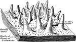
Mucous Membrane from the Jejunum
The mucous membrane from the jejunum. Labels: 1, Villi (folds of lining mucous membrane) in miniature.…
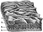
A Portion of the Mucous Membrane from the Small Intestine
Portion of the mucous membrane from the small intestine, magnified, showing the villi on its free surface,…
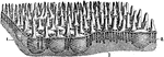
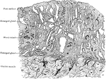
Mucous Membrane of the Small Intestine
Free surface of the mucous membrane of the small intestine, showing villi, solitary glands, and opening…
Mucous Membrane of Uterus in Fourth Month of Pregnancy
Section of mucous membrane lining body of uterus (decidua vera); fourth month of pregnancy.
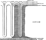
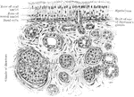
Gastropulmonary Mucous Membrane
Diagram of the gastropulmonary mucous membrane, showing the continuity of all its parts.
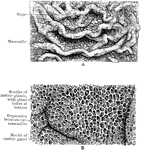
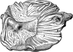
Stomach Mucous Membrane
The mucous membrane of the stomach. A, Natural size. B. Magnified. In A the rugae and the mammilated…
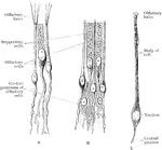
Olfactory and Supporting Cells
Olfactory and supporting cells in a frog and a human. A. Frog. B. Human. C. Human.
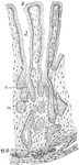
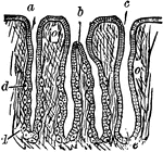
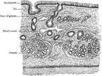
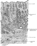
Pyloric Gland
Section showing the pyloric glands. Labels: s, free surface; d, ducts of pyloric glands; n, neck of…
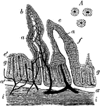
Rabbit's Intestinal Mucous Membrane
Vertical section of the intestinal mucous membrane of the rabbit. Two villi are represented, in one…
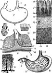
Horse Stomach
Internal aspect of a horse stomach, opened from below. Labels: a, cuticular mucous membrane; b, villous…
Views of the Stomach
Views of the stomach. Labels: A. stomach (human). B. Same, anterior wall removed. C. Portion of stomach,…