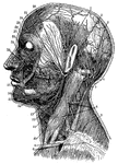
"Superficial arteries and nerves of the face and neck. 1, Temporal artery; 2, artery behind the ear; 3, occipital artery; 4, greater occipital nerve; 5, smaller occipital nerve; 6, nerve of the neck; 7, trapezius muscle; 8, clavicular nerves; 9, clavicle; 10, sterno-cleido-mastoid muscle; 11, outer artery of the head; 12, inner artery of the head; 13, salivary gland; 14, nerves of the lower jaw; 15, outer maxillary artery; 16, nerve of the chin; 17, circular muscle of the mouth; 18, greater yoke muscle; 19, nerves below the eye; 20, masseter, or chewing muscle; 21, ear passage; 22, arteries of the forehead; 23; nerves of the forehead; 24, eye-closing muscle; 25, facial artery; 26, facial nerve." -Foster, 1921

Arteries and Nerves

"Superficial arteries and nerves of the face and neck. 1, Temporal artery; 2, artery behind the ear;…

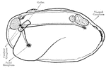
The nervous system of a clam.

Clam

The nervous system of a clam.

The nerves of the finger.

Finger

The nerves of the finger.

"Nerves of the had. 1, Nerves of the skin; 2, tendons; 3, arteries of the palm of the hand; 4, elbow nerve; 5, elbow artery; 6, nerve of the forearm; 7, nerve of the under-arm; 8, artery of the under-arm." -Foster, 1921

Hand Nerves

"Nerves of the had. 1, Nerves of the skin; 2, tendons; 3, arteries of the palm of the hand; 4, elbow…

This illustration shows nerve cells from spinal ganglia.

Nerve Cells from Spinal Ganglia

This illustration shows nerve cells from spinal ganglia.

Nerve fibers. Labels: a, nerve-fiber, showing complete interruption of the white substance; b, another nerve fiber with nucleus. In both these nerve fibers the white substance is stained black with osmic acid, and the axon is seen running as an uninterrupted strand through the center of the fiber. c, ordinary nerve fiber unstained; d, e, smaller nerve fiber; f, varicose nerve fiber; g, non-medullated nerve fibers.

Nerve Fibers

Nerve fibers. Labels: a, nerve-fiber, showing complete interruption of the white substance; b, another…

Nerve Ganglia, or Knots (sing. Ganglion; Knot) occur as collections of nerve cells on the course of a nerve, forming and obvious swelling. In surgery, it is the swelling caused by the collection of fluid in the fibrous sheath which surrounds a tendon. It arises from inflammation of the tendon-sheath, and occurs most often at the back of the wrist. Sometimes the cyst may be broken by firm pressure or by a smart blow, and the fluid be thus dispersed; or a puncture may be made, with proper antiseptic precautions, and the fluid be pressed out. Sometimes counter-irritation with iodine causes absorption. In any case, firm pressure with pad and bandage must be used afterwards, to prevent refilling until the cyst walls have united. This illustration shows a cross-section of a spinal cord and spinal nerve cells (g being ganglia).

Nerve Ganglia (Spinal)

Nerve Ganglia, or Knots (sing. Ganglion; Knot) occur as collections of nerve cells on the course of…

Diagram to indicate the arrangement of the Sympathetic Nervous Centers and Fibers.

Nervous Centers

Diagram to indicate the arrangement of the Sympathetic Nervous Centers and Fibers.

Part of the nervous system viewed from in front.

Nervous System

Part of the nervous system viewed from in front.

Diagram illustrating the general arrangement of the cerebrospinal nervous system.

Diagram of the Human Nervous System

Diagram illustrating the general arrangement of the cerebrospinal nervous system.

Diagram of nervous system. Labels: a, a, cortex of cerebral hemispheres; b, b, cell body and dendrites of upper motor neuron, situated in cerebral cortex; b', axon of upper motor neuron, branching at its termination near the dendrites of lower motor neuron, situated in the ventral horn of gray matter in the spinal cord; B', axon of lower motor neuron passing to its termination in a voluntary muscle fiber B"; C, cell body and dendrites of supper sensory neuron, situated in the medulla oblongata; C'C', axons of upper sensory neurons, terminating in cortex; c, cell body of lower sensory neuron situated in the dorsal root ganglion; c''', dendrite of lower motor neuron, conducting impulses from the periphery to the central nervous system; c", long axon of lower sensory neuron, conducting impulses toward the brain; c', short axon of lower sensory neuron, conducting impulses direct to ventral horn. (For the sake of simplicity the connection with the cerebellum are omitted.)

Nervous System Diagram

Diagram of nervous system. Labels: a, a, cortex of cerebral hemispheres; b, b, cell body and dendrites…

Diagram of a neuron. Labels: A, axon arising from the cell-body and branching at its termination; D, dendrites; C and N, cell-body composed of C, cytoplasm, and N, nucleus.

Diagram of a Neuron

Diagram of a neuron. Labels: A, axon arising from the cell-body and branching at its termination; D,…

"Ideal plan of the double-ringed body of a vertebrate. N, neural canal; H, haemal canal; the body separating them is the centrum of any vertebra, bearing e, and epapophysis, and y, a hypapophysis; n, n, neurapophyses; d, d, diapophyses; ns, bifid neural spine; pl, pl, pleurapophyses; h, h, haemapophyses; hs, bifid haemal spine. Drawn by Dr. R. W. Shufeldt, USA, After Owen. The Axial Skeleton of a bird or any vertebrated animal, that is, one having a back-bone, exhibits in cross-section two rings or hoops, one above and the other below a central point, like the upper and lower loops of a figure 8. The upper ring is the neural arch, so called because such cylinder encloses a section of the cerebro-spinal axis, or principal nervous system of a vertebrate (brain and spinal cord, whence arise all the nerves of the body, excepting those of the sympathetic nervous system). The lower ring is the haema arch, which similarly contains a section of the principal blood vessals and viscera.

Axial Skeleton

"Ideal plan of the double-ringed body of a vertebrate. N, neural canal; H, haemal canal; the body separating…