Diagram of fetal circulation.

Diagram of Fetal Circulation

Diagram of fetal circulation.

A human embryo of the fourth week. I, the chorion; 3, part of the amnion; 4, umbilical vesicle with its long pedicle passing into the abdomen; 7, the heart; 8, the liver; 9, the visceral arch destined to form the lower jaw, beneath which are two other visceral arches separated by the branchial clefts; 10, rudiment of the upper extremity, 11, that of the lower extremity; 12, the umbilical cord; 15, the eye; 16, the ear; 17, cerebral hemispheres; 18, optic lobes, corpora quadrigemina.

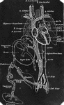
Embryo at Fourth Week

A human embryo of the fourth week. I, the chorion; 3, part of the amnion; 4, umbilical vesicle with…

Human embryo of fifth week with umbilical vesicle. The human umbilical vesicle never exceeds the size of a small pea.

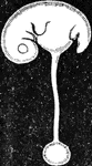
Embryo of Fifth Week

Human embryo of fifth week with umbilical vesicle. The human umbilical vesicle never exceeds the size…
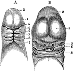
A, Magnified view of the head and neck of a human embryo of three weeks. Labels: 1, anterior cerebral vesicle or cerebrum; 2, middle cerebral vesicle; 3, middle or frontonasal process; 4, superior maxillary process, or first visceral arch, and below it the first cleft; 7, 8, 9, second, third, and fourth arches and clefts. B, Anterior view of the head of a human fetus of about the fifth week. 1, 2, 3, 5, the same parts as in A; 4, the external nasal or lateral frontal process: 6, the superior maxillary process; 7, the lower jaw; X, the tongue; 8, first branchial cleft becoming the meatus auditorius externus.

Head of an Embryo

A, Magnified view of the head and neck of a human embryo of three weeks. Labels: 1, anterior cerebral…

Diagram of an early stage of the formation of the human placenta. Labels: a, embryo; b, amnion; c, placental vessels; d, decidua reflexa; e, allantois; f, placental villi; g, mucous membrane.

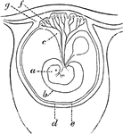
Early Formation of the Placenta

Diagram of an early stage of the formation of the human placenta. Labels: a, embryo; b, amnion; c, placental…

Diagrammatic view of a vertical transverse section of the uterus at the seventh week of pregnancy. Labels: c, c', cavity of uterus, which becomes the cavity of the decidua, opening at c, the cornua, into the Fallopian tubes, and at c' into the cavity of the cervix, which is closed by a plug of mucus; dv, decidua vera; dr, decidua reflexa, with the sparser villi imbedded in its substance; ds, decidua serotina, involving the more developed chorionic villi of the commencing placenta. The fetus is seen lying in the amniotic sac; passing up from the umbilicus is seen the umbilical pedicle of the yolk sac, which lies in the cavity between the amnion and chorion.

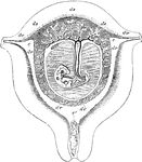
Uterus at Seventh Week of Pregnancy

Diagrammatic view of a vertical transverse section of the uterus at the seventh week of pregnancy. Labels:…