This illustration shows a representation of the circulation of the blood, in its essential features. The arrows indicate the course of the blood. a, arteries; aur., auricle or receiving portion of the heart; d, digestive tract; c. d., capillaries of the digestive tract; c.r., capillaries of the respiratory organs; c.s., capillaries of the system; va., valves; ve, veins; vt, ventricle.

Blood Circulation

This illustration shows a representation of the circulation of the blood, in its essential features.…

Transverse section of the chest.

Transverse Section of the Chest

Transverse section of the chest.

This illustration shows the thorax of a crayfish with a portion of the carapace removed to show the gills.

Gills (Crayfish)

This illustration shows the thorax of a crayfish with a portion of the carapace removed to show the…
"<em>A</em>, division of left bronchus to upper lobe; <em>B</em>, left branch of the pulmonary artery; <em>C</em>, left bronchus; <em>D</em>, left superior pulmonary vein; <em>E</em>, left inferior pulmonary vein; <em>F</em>, left auricle; <em>K</em>, inferior vena cava; <em>L</em>; division of the right bronchus to lower lobe; <em>M</em>, right inferior pulmonary vein; <em>N</em>, right superior pulmonary vein, <em>O</em>, right branch of the pulmonary artery; <em>P</em>, division of right bronchus to upper lobe; <em>R</em>, left ventricle; <em>S</em>, right ventricle." &mdash; Blaisedell, 1904

Lungs

"A, division of left bronchus to upper lobe; B, left branch of the pulmonary artery;…

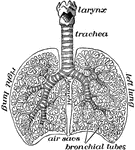
"The lungs fill up most of the cavity of the chest. One lies on either side of the heart which is in the middle of the chest. The lungs in animals are called lights because they are spongy sacs and so light as to float when thrown into water." &mdash;Davison, 1910

The lungs

"The lungs fill up most of the cavity of the chest. One lies on either side of the heart which is in…

<i>Fusus antiquus</i>. "...a division of prosobranchiate gastropods, having the lip of the shell notched, canaliculate, or tubular, for the protrusion of a respiratory siphon. a, branchial siphon; b, proboscis; c, operculum; d, d, tentacles; f, foot." &mdash;Whitney, 1889

Red Whelk

Fusus antiquus. "...a division of prosobranchiate gastropods, having the lip of the shell notched, canaliculate,…

Outline showing the general form of the larynx, trachea, and bronchi, as seen from behind. Labels: h, the great cornu of the hyoid bone; e,epiglottis; t, superior cornu of the thyroid cartilage ; t1, inferior cornu of the thyroid cartilage; a, points to the back of both the arytenoid cartilages, which are surmounted by the cornicula; c, the middle ridge on the back of the cricoid cartilage; tr, the posterior membranous part of the trachea; b, b1, right and left bronchi.

Back View of Respiratory Apparatus

Outline showing the general form of the larynx, trachea, and bronchi, as seen from behind. Labels: h,…

Outline showing the general form of the larynx, trachea, and bronchi, as seen from front. Labels: h, the great cornu of the hyoid bone; e,epiglottis; t, superior and inferior cornu of the thyroid cartilage; c, middle of the cricoid cartilage; tr, the trachea, showing sixteen cartilaginous rings; b, the right bronchus; b1, the left bronchus.

Front View of Respiratory Apparatus

Outline showing the general form of the larynx, trachea, and bronchi, as seen from front. Labels: h,…
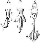
The development of the respiratory organs. A, is the esophagus of a chick on the fourth day of incubation, with the rudiments of the trachea on the lung of the left side, viewed laterally; 1, the inferior wall of the esophagus; 2, the upper portion of the same tube; 3, the rudimentary lung; 4, the stomach. B, is the same object seen from below, so that both lungs are visible. C, shows the tongue and respiratory organs of the embryo of a horse; 1, the tongue; 2, the larynx; 3, the trachea; 4, the lungs, viewed from the upper side.

Development of Respiratory Organs

The development of the respiratory organs. A, is the esophagus of a chick on the fourth day of incubation,…
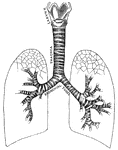
"Larynx, trachea, and bronchi, showing the manner of division, and the rings of cartilage." &mdash; Tracy, 1888

Respiratory system

"Larynx, trachea, and bronchi, showing the manner of division, and the rings of cartilage." —…
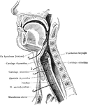
Operative approaches through the front of the neck to the larynx, pharynx, and trachea. a: Approach to pharynx between the hyoid and the thyroid cartilages. b: Approach to the larynx between the thyroid and cricoid cartilages. c: approach to the larynx below the cricoid cartilage and above the isthmus of the thyroid gland. d: Approach for low tracheotomy.

Upper Respiratory Tract

Operative approaches through the front of the neck to the larynx, pharynx, and trachea. a: Approach…
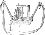
The stethograph or pneumograph consists of a thick rubber of elliptical shape about three inches long, to one end of which a rigid gutta-percha tube is attached. Labels: h, tambour fixed at right angles to plate of steel f; c and d, arms by which instrument is attached to chest by belt e. When the chest expands, the arms are pulled asunder, which bends the steel plate, and the tambour is affected by the pressure of b, which is attached to it on the one hand, and to the upright in connection with horizontal screw g.

Stethograph or Pneumograph

The stethograph or pneumograph consists of a thick rubber of elliptical shape about three inches long,…
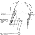
The stethometer consists of a frame formed of two parallel steel bars joined by a third at one end. At the free end of the bars is attached a leather strap, by means of which the apparatus may be suspended from the neck. Attached to the inner end of one bar is a tambour and ivory button, to the end of the other an ivory button. When in use, the apparatus is suspended with the transverse bar posteriorly, the button of the tambour is placed on the part of the chest the movement of which it is desire to record, and the other button is made to press upon the corresponding side of the chest, so that the chest is held between a pair of callipers.

Stethometer

The stethometer consists of a frame formed of two parallel steel bars joined by a third at one end.…

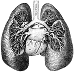
"Larynx, Trachea, and the Bronchi. (Front view.) <em>A</em>, epiglottis; <em>B</em>, thyroid cartilage; <em>C</em>, cricothyroid membrane, connecting with the cricoid cartilage below, all forming the larynx; <em>D</em>, rings of the trachea." &mdash; Blaisedell, 1904

Trachea

"Larynx, Trachea, and the Bronchi. (Front view.) A, epiglottis; B, thyroid cartilage;…