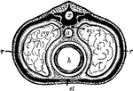
A Diagrammatic section across the body in the chest region. Labels: x, the dorsal tube, which contains the spinal cord; the black mass surrounding it is a vertebra; a, the gullet (esophagus), a part of the alimentary canal; h, the heart; sy, sympathetic nervous system; ll, lungs; the dotted lines around them are the pleurae; rr, ribs; st, the breastbone.

Section Across the Body in the Chest Region

A Diagrammatic section across the body in the chest region. Labels: x, the dorsal tube, which contains…
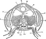
"Diagrammatic cross-section of Cray-fish in the thoracic region, to show relation of circulation and respiration. a, appendage; c, carapace; c.f., flap of carapace overhanging the gills; d, digestive tube; g, gill; h, heart; l, liver; m, body muscles; m', muscles of the appendages; n.c., nerve cord; p.s., paricardial sinus; r, reproductive glands; st, sternal artery; v.a., ventral artery; v.s., ventral blood sinus in which the nerve cord lies." — Galloway

Cray-fish

"Diagrammatic cross-section of Cray-fish in the thoracic region, to show relation of circulation and…

The eleventh and twelfth ribs have each a single articular facet on the head, which is of rather large size; they have no neck or tuberosity, and are pointed at the extremity.

Eleventh Rib

The eleventh and twelfth ribs have each a single articular facet on the head, which is of rather large…
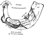
The first rib is the shortest and the most curved of all the ribs; it is broad and flat, its surfaces looking upward and downward, and its borders inward and outward. The head is of small size, rounded, and presents only a single articular facet for articulation with the body of the first thoracic vertebra.

First Rib

The first rib is the shortest and the most curved of all the ribs; it is broad and flat, its surfaces…

"Galeodes sp., one of the Solifugae. Ventral view to show legs and somites. I to VI, The six leg-bearing somites of the prosoma. opisth I, First or genital somite of the opisthosoma. ge, Site of the genital aperture. st, Thoracic tracheal aperture. l2, Anterior tracheal aperture of the opisthosoma in somite 2 of the opisthosoma. l3, Tracheal aperture in somite 3 of the opisthosoma. a, Anus." — The Encyclopedia Britannica, 1910

Galeodes

"Galeodes sp., one of the Solifugae. Ventral view to show legs and somites. I to VI, The six leg-bearing…

"The lymph vessels of the body. rc, the thoracic duct; lac, the lacteals taking the lymph and fatty part of food from the intestines." —Davison, 1910

Lymph Vessels

"The lymph vessels of the body. rc, the thoracic duct; lac, the lacteals taking the lymph and fatty…
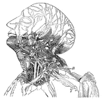
This diagram shows the lumphatics of the head and neck. It shows the glands, and B, the thoracic duct as it empties into the left innominate vein at the junction of the left jugular and subclavian veins.

Lymphatics

This diagram shows the lumphatics of the head and neck. It shows the glands, and B, the thoracic duct…

"Mullus barbatus (Red Mullet), with thoracic ventral fins." — Encyclopedia Britannica, 1893

Red Mullet

"Mullus barbatus (Red Mullet), with thoracic ventral fins." — Encyclopedia Britannica, 1893

"The Cervical and Thoracic Portions of the Sympathetic Nerve and their Main Branches. In the center of the figure, running almost vertically, is shown the right pneumogastrix nerve. To the left may be seen a chain of ganglia of the sympathetic nerve, running along the vertebrae. The distribution of some smaller ganglia and nerve branches in the neck and chest regions are also shown." — Blaisedell, 1904

Sympathetic nerve

"The Cervical and Thoracic Portions of the Sympathetic Nerve and their Main Branches. In the center…

The second rib is much longer than the first, but bears a very considerable resemblance to it in the direction of its curvature. The nonarticular portionof the tuberosity is occasionally only slightly marked.

Second Rib

The second rib is much longer than the first, but bears a very considerable resemblance to it in the…

Anterior and posterior view of adult spine.

Anterior and Posterior Views of Spine

Anterior and posterior view of adult spine.

Lateral view of adult spine.

Lateral View of Spine

Lateral view of adult spine.

The tenth rib has only a single articular facet on its head.

Tenth Rib

The tenth rib has only a single articular facet on its head.

Diagram showing the position of the thoracic and abdominal organs. labels: 1, lower border of right lung; 2, the same of the left lung; 3, liver, right lobe; 4, liver, left lobe; 5, suspensory ligament of the liver; 6, fundus of gall-bladder; 7, cardia of stomach; 8, fundus of stomach; 9, lower border of stomach; 10, position of pylorus; 11, caecum; 12, vermiform appendix; 13, ascending colon; 14, right flexure of colon; 15, transverse colon, concealed by, 19, convolutions of the small intestine; 20, termination of ileum, ascending from right to left; 21, bladder, distended, partly covered by peritoneum; 22, the part of the bladder which is not covered by peritoneum.

Section Across the Forearm

Diagram showing the position of the thoracic and abdominal organs. labels: 1, lower border of right…
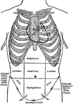
Diagram of the thoracic and abdominal regions. Labels: A, aortic valve; P, pulmonary valve; M, mitral valve; T, tricuspid valve.

Thoracic and Abdominal Regions

Diagram of the thoracic and abdominal regions. Labels: A, aortic valve; P, pulmonary valve; M, mitral…

"The three branches from left to right are the unnamed ones. The primitive left carotid and the subclavian left one. The small branch in the cure is the bronchial one." — Richardson, 1906

Thoracic Aorta

"The three branches from left to right are the unnamed ones. The primitive left carotid and the subclavian…
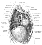
Deep structures of the right thoracic cavity, after removal of the right lung.

Thoracic Cavity after Removal of Lung

Deep structures of the right thoracic cavity, after removal of the right lung.

"The lacteals conduct the chyle from the intestines into numerous glands nearby, called the mesenteries, from which a fewer number of lacteals lead the chyle into one large tube in front of the spinal column, called the thoracic duct." —Brown, 1884

Thoracic duct and lacteals

"The lacteals conduct the chyle from the intestines into numerous glands nearby, called the mesenteries,…
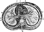
"The transverse section of the thorax. 1, anterior mediastinum; 2, internal mammary vessels; 3, triangularis sterni; 4, 5, phrenic nerves; 6, thoracic duct; 7, esophagus; 8, vena azygos major; 9, thoracic aorta; 19, sympathetic; R. V., right ventricle; R. A., right auricle; P.A., pulmonary artery; A., aorta; C., superior vena cava; V., dorsal vertebra." — Richardson, 1906

Thorax

"The transverse section of the thorax. 1, anterior mediastinum; 2, internal mammary vessels; 3, triangularis…
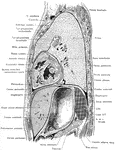
Lateral, sagittal section through the left thorax and upper portion of abdomen, viewed from the left. The plane of the section lies 4 cm to the left of the median plane.

Side View of the Thorax and Part of the Abdomen

Lateral, sagittal section through the left thorax and upper portion of abdomen, viewed from the left.…

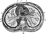
Frontal section through the thorax, passing through the mediastinum and the middle of the humeral heads.
Frontal Section Through the Thorax

Frontal section through the thorax, passing through the mediastinum and the middle of the humeral heads.

Transverse section of the thorax. Labels: 1, anterior mediastinum; 2, internal mammary vessels; 3, triangularis sterni; 4, 5, phrenic nerves; 6, thoracic duct; 7, esophagus; 8, vena azygos major; 9, thoracic aorta; 10, sympathetic; R. V., right ventricle; R. A., right auricle; P. A. pulmonary artery; A., aorta; C., superior vena cava; V., dorsal vertebra.

Transverse Section of the Thorax

Transverse section of the thorax. Labels: 1, anterior mediastinum; 2, internal mammary vessels; 3, triangularis…

"Triarthrus Becki, Green. a, Restored thoracic limbs in transverse section of the animal; b, section across a posterior somite; c, section across one of the sub-terminal somites." — The Encyclopedia Britannica, 1910

Triarthrus

"Triarthrus Becki, Green. a, Restored thoracic limbs in transverse section of the animal; b, section…
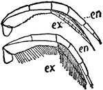
"Triarthrus Becki, Green. Dorsal view of second thoracic leg with and without setae. en, Inner ramus; ex, Outer ramus." — The Encyclopedia Britannica, 1910

Triarthrus

"Triarthrus Becki, Green. Dorsal view of second thoracic leg with and without setae. en, Inner ramus;…
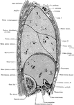
Sagittal section through the trunk, 6 cm to the right of the median plane, viewed from the right. Note the close proximity of the duodenum and colon to the right kidney.

Side View of the Trunk

Sagittal section through the trunk, 6 cm to the right of the median plane, viewed from the right. Note…

The eleventh and twelfth ribs have each a single articular facet on the head, which is of rather large size; they have no neck or tuberosity, and are pointed at the extremity.

Twelfth Rib

The eleventh and twelfth ribs have each a single articular facet on the head, which is of rather large…

The spinal column, right lateral view and dorsal view.

Lateral and Dorsal View of the Vertebral Column

The spinal column, right lateral view and dorsal view.