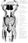
The passage to the esophagus and windpipe. Labels: c, the tongue; d, the soft palate ending in g, the uvula; h, the epiglottis; i, the glottis; I, the esophagus; f, the pharynx.

Passage to the Esophagus and Windpipe

The passage to the esophagus and windpipe. Labels: c, the tongue; d, the soft palate ending in g, the…
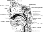
Anterior portion of mesial sagittal section of child's head, probably of about three year.

Mesial Sagittal Section of Child's Face

Anterior portion of mesial sagittal section of child's head, probably of about three year.
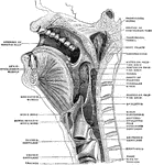
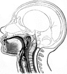
Sagittal median section of the head and neck. The head is thrown backward into complete extension which explains the relations between the lower jaw and the hyoid bone as seen in the figure.

Sagittal Section of the Head and Neck

Sagittal median section of the head and neck. The head is thrown backward into complete extension which…
Section of the head through the point of chin.

Cross Section of Head Through the Chin

Section of the head through the point of chin.

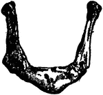
The hyoid, os hyoides, or tongue bone, is an isolated, U-shaped bone lying in front of the throat, just above the Adam's apple; it supports the tongue, and give attachment to some of its numerous muscles.

Human Hyoid Bone

The hyoid, os hyoides, or tongue bone, is an isolated, U-shaped bone lying in front of the throat, just…
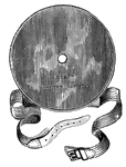
This part of the laryngoscope consists of a small mirror fixed to a long handle.

Laryngoscope

This part of the laryngoscope consists of a small mirror fixed to a long handle.

This part of the Laryngoscope consists of a large concave mirror with a small hold in the middle.

Laryngoscope

This part of the Laryngoscope consists of a large concave mirror with a small hold in the middle.
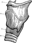
External view of the left side of Larynx. 1: Front portion of hyoid bone; 2: Upper edge of larynx; 3: Lower portion of larynx; 4: Second ring of trachea.

Larynx

External view of the left side of Larynx. 1: Front portion of hyoid bone; 2: Upper edge of larynx; 3:…
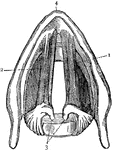
Cross section of the larynx above the vocal cords. 1: Right vocal cord. 2: Left vocal cord. 3: Cartilages to which the vocal cords are attached behind; 4: Front edge of the larynx.

Larynx

Cross section of the larynx above the vocal cords. 1: Right vocal cord. 2: Left vocal cord. 3: Cartilages…

Muscles of the larynx. Side view. Right ala of thyroid cartilage removed.

Side View of the Muscles of the Larynx

Muscles of the larynx. Side view. Right ala of thyroid cartilage removed.
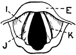
This illustration shows a vertical section of the larynx and its many parts (A. Thyroid Cartilage; B. Cricoid Cartilage; C. hyoid bone; D. Rings of trachea; E. Epiglottis; F. Arytenoid Cartilage; G. Thyro-arytenoid muscle; H. Adam's Apple; I. False Vocal Cords; J. True Vocal Cords; K. Ventricle; L. Rima Glottis).

Vertical Section of Larynx

This illustration shows a vertical section of the larynx and its many parts (A. Thyroid Cartilage; B.…
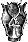
Larynx, viewed from above.

Larynx Viewed from Above

Larynx, viewed from above.

Labels: T, thyroid cartilage: C, cricoid cartilage; Tr, trachea; H, hyoid bone; E, epiglottis; I, joint of thyroid cartilage; Ar, arytenoid cartilages.

Back View of the Larynx

Labels: T, thyroid cartilage: C, cricoid cartilage; Tr, trachea; H, hyoid bone; E, epiglottis; I, joint…

Muscles of the larynx, back view.

Back View of the Muscles of the Larynx

Muscles of the larynx, back view.

Coronal section of the larynx, rear view of front half.

Coronal Section of the Larynx

Coronal section of the larynx, rear view of front half.

Coronal section through larynx, to show its three compartments.

Coronal Section Through Larynx

Coronal section through larynx, to show its three compartments.

"Cartilages and Ligaments of the Larynx. (Front view.) <em>A</em>, hyoid bone; <em>B</em>, membrane attached to hyoid bone and the shield-shaped cartilage below (thyroid); edges of this shield-shaped cartilage unite at <em>C</em> (Adam's apple is the V-shaped groove on a line with <em>B</em> and <em>C</em>; <em>D</em>, membrane between the shield-shaped cartilage and the signet-ring cartilage below; <em>E</em> cricoid, or signet-ring, cartilage; <em>F</em>, upper ring of the windpipe." &mdash; Blaisedell, 1904

Front view of the larynx

"Cartilages and Ligaments of the Larynx. (Front view.) A, hyoid bone; B, membrane…
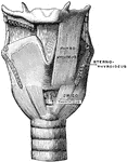
Muscles of the larynx, front view. The Sternothyroid and right Thyrohyoid have been removed.

Front View of the Muscles of the Larynx

Muscles of the larynx, front view. The Sternothyroid and right Thyrohyoid have been removed.
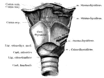
The part of the respiratory tract between the pharyna and the trachea. Responsible for speech.

Human Larynx

The part of the respiratory tract between the pharyna and the trachea. Responsible for speech.
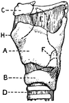
This illustration shows a lateral aspect of the larynx and its multiple parts (A. Thyroid Cartilage; B. Cricoid Cartilage; C. hyoid bone; D. Rings of trachea; E. Epiglottis; F. Arytenoid Cartilage; G. Thyro-arytenoid muscle; H. Adam's Apple; I. False Vocal Cords; J. True Vocal Cords; K. Ventricle; L. Rima Glottis).

Lateral Aspect of Larynx

This illustration shows a lateral aspect of the larynx and its multiple parts (A. Thyroid Cartilage;…

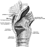
Mesial section through larynx, to show the outer wall of the right half.

Mesial Section Through Larynx

Mesial section through larynx, to show the outer wall of the right half.

"Cartilages and Ligaments of the Larynx. (Front view.) <em>A</em>, epiglottis; <em>B</em>, thyroid cartilage; <em>C</em>, ladle-shaped cartilage; <em>E</em>, cricoid cartilage; <em>F</em>, upper ring of the windpipe." &mdash; Blaisedell, 1904

Posterior view of the larynx

"Cartilages and Ligaments of the Larynx. (Front view.) A, epiglottis; B, thyroid cartilage;…

Sagittal section of the larynx, right half.

Sagittal Section of the Larynx

Sagittal section of the larynx, right half.

Labels: T, thyroid cartilage: C, cricoid cartilage; Tr, trachea; H, hyoid bone; E, epiglottis; I, joint of thyroid cartilage.

Side View of the Larynx

Labels: T, thyroid cartilage: C, cricoid cartilage; Tr, trachea; H, hyoid bone; E, epiglottis; I, joint…
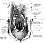
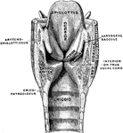
Superior aperture of larynx, exposed by laying open the pharynx from behind.

Superior Aperture of Larynx

Superior aperture of larynx, exposed by laying open the pharynx from behind.
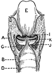
This illustration shows a longitudinal section of  the larynx as seen from behind (A. Thyroid Cartilage; B. Cricoid Cartilage; C. hyoid bone; D. Rings of trachea; E. Epiglottis; F. Arytenoid Cartilage; G. Thyro-arytenoid muscle; H. Adam's Apple; I. False Vocal Cords; J. True Vocal Cords; K. Ventricle; L. Rima Glottis).

Longitudinal Section of Larynx Seen from Behind

This illustration shows a longitudinal section of the larynx as seen from behind (A. Thyroid Cartilage;…
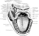
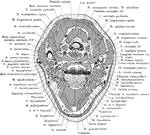
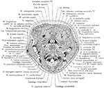
Horizontal section through mouth and pharynx at the level of the tonsils.

Horizontal Section Through Mouth and Pharynx

Horizontal section through mouth and pharynx at the level of the tonsils.

The mouth, nose, and pharynx, with the commencement of gullet (esophagus) and larynx, as exposed by a section a little to the left of the median plane of the head. Labels: a, vertebrae; b, gullet; c, trachea; d, larynx; e, epiglottis; f, soft palate; g, opening of Eustachian tube; k, tongue; l, hard palate; o, p, q, inferior turbinate bones of left nasal chamber.

The Mouth, Nose, and Pharynx

The mouth, nose, and pharynx, with the commencement of gullet (esophagus) and larynx, as exposed by…

The mouth, nose, and pharynx, with the larynx and commencement of gullet (esophagus), seen in section. Labels: a, vertebral column; b, gullet (esophagus); c, trachea; d, larynx; e, epiglottis; f, soft palate, between f and e is the opening at back of cavity or faces; g, opening of Eustachian tube; h, nasal cavity; k, tongue; l, hard palate; m, sphenoid bone at base of skull; n, roof of nasal cavity; o, p, q, placed in nasal cavity.

The Mouth, Nose, and Pharynx

The mouth, nose, and pharynx, with the larynx and commencement of gullet (esophagus), seen in section.…

The mouth, nose, and pharynx, with the commencement of the gullet (esophagus) and larynx, as exposed by a section, a little to the left of the median plane of the head. Labels: a, vertebral column; b, gullet; c, windpipe; d, larynx; e, epiglottis; f, soft palate; g, opening of Esutachian tube; k, tongue; l, hard palate; m; the sphenoid bone on the base of the skull; n, the fore part of the cranial cavity; o,p,q, the turbinate bones of the outer side of the left nostril chamber.

The Mouth, Nose, and Pharynx

The mouth, nose, and pharynx, with the commencement of the gullet (esophagus) and larynx, as exposed…

"Diagram of a Sectional View of Nasal and Throat Passageways. <em>C</em>, nasal cavities; <em>T</em>, tongue; <em>L</em>, lower jaw; <em>M</em>, Mouth; <em>U</em>, uvula; <em>E</em>, epiglottis; <em>G</em>, larynx; <em>O</em>, gullet, or esophagus." &mdash; Blaisedell, 1904

Nasal and throat passageways

"Diagram of a Sectional View of Nasal and Throat Passageways. C, nasal cavities; T,…

Anterior part of section across neck at level of false vocal cords; on left side ventricle of larynx is exposed.

Anterior Part of Section Across Neck at False Vocal Cords

Anterior part of section across neck at level of false vocal cords; on left side ventricle of larynx…

Anterior part of section across neck at level of fourth cervical vertebra, passing through upper part of superior aperture of larynx.

Anterior Part of Section Across Neck at Fourth Cervical Vertebra

Anterior part of section across neck at level of fourth cervical vertebra, passing through upper part…

Modes of approach to the esophagus and the cervical sympathetic ganglion shown by means of a cross section of the neck at the level of the 7th cervical vertebra. A: Approach to esophagus internal to carotid sheath. B : An incision in midline beneath the hyoid bone to the esophagus. C: An approach similar to (A), median to the carotid sheath, and (D), external to the carotid sheath, to expose the cervical sympathetic ganglion and trunk and the inferior, thyroid artery.

Cross Section of Neck at the 7th Cervical Vertebra

Modes of approach to the esophagus and the cervical sympathetic ganglion shown by means of a cross section…
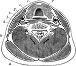
Section of the neck through larynx.

Cross Section of Neck Through the Larynx

Section of the neck through larynx.

Transverse section through the lower part of the neck, to show the arrangement of the cervical fascia. Labels: a, Trapezius; b, sternomastoid; c, depressors of hyoid bone; d, platysma; e, anterior spinal muscles; f, scalenus anticus; g, carotid artery; h, external jugular vein; i, posterior spinal muscles; T, trachea, with gullet behind and thyroid body in front.

Transverse Section of the Neck

Transverse section through the lower part of the neck, to show the arrangement of the cervical fascia.…
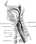
The anterior all of the pharynx with its orifices, seen from behind.

Pharynx Wall

The anterior all of the pharynx with its orifices, seen from behind.

Operative approaches through the front of the neck to the larynx, pharynx, and trachea. a: Approach to pharynx between the hyoid and the thyroid cartilages. b: Approach to the larynx between the thyroid and cricoid cartilages. c: approach to the larynx below the cricoid cartilage and above the isthmus of the thyroid gland. d: Approach for low tracheotomy.

Upper Respiratory Tract

Operative approaches through the front of the neck to the larynx, pharynx, and trachea. a: Approach…
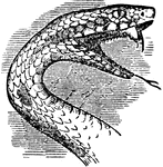
"The prey of a serpent is oven thicker than the serpent itself, and to admit of its being swallowed, the throat and body are very dilatable. The bones of the head are adapted to the necessity of a great expansion of the mouth and dilation of the throat." &mdash; Chambers, 1881

Serpent Head

"The prey of a serpent is oven thicker than the serpent itself, and to admit of its being swallowed,…
The red-throated tamatia, native to Guiana.

Red-Throated Tamatia

The red-throated tamatia, native to Guiana.

The trachea and bronchi. The thyroid body is indicated by a dotted line.

Trachea and Bronchi

The trachea and bronchi. The thyroid body is indicated by a dotted line.

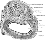
Transverse section of trachea and esophagus of child, seen from below.

Transverse Section of Trachea and Esophagus

Transverse section of trachea and esophagus of child, seen from below.
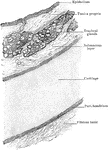
Transverse section of trachea, showing general arrangement of its wall.

Transverse Section of Trachea Showing Arrangement of Walls

Transverse section of trachea, showing general arrangement of its wall.
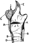
"<em>A</em>, epiglottis; <em>B</em>, section of hyoid bone; <em>C</em>, false vocal cords which have nothing to do with the production of voice; <em>D</em>, oblong opening between the false and true vocal cords; <em>E</em>, true vocal cord;<em>F</em>, section of the thyroid cartilage; <em>H</em>, section of the anterior portion of the cricoid cartilage; <em>K</em>, trachea; <em>L</em>, section of the posterior portion of the cricoid cartilage; <em>M</em>, ladle-shaped cartilage." &mdash; Blaisedell, 1904

Vocal cords

"A, epiglottis; B, section of hyoid bone; C, false vocal cords which have…

This illustration shows the vocal cords as seen from above during phonation (A. Thyroid Cartilage; B. Cricoid Cartilage; C. hyoid bone; D. Rings of trachea; E. Epiglottis; F. Arytenoid Cartilage; G. Thyro-arytenoid muscle; H. Adam's Apple; I. False Vocal Cords; J. True Vocal Cords; K. Ventricle; L. Rima Glottis).

Vocal Cords Seen from above During Phonation

This illustration shows the vocal cords as seen from above during phonation (A. Thyroid Cartilage; B.…

The Yellow-throated Vireo, Vireo flavifrons, is a small American songbird. Adults are mainly olive on the head and upperparts with a yellow throat and white belly; they have dark eyes with yellow "spectacles". The tail and wings are dark with white wing bars. They have thick blue-grey legs and a stout bill. Their breeding habitat is open deciduous woods in southern Canada and the eastern United States

Yellow-throated Vireo

The Yellow-throated Vireo, Vireo flavifrons, is a small American songbird. Adults are mainly olive on…