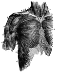
A frontal view of the chest muscles. 1, sterno-hyoid; 2, sterno-mastoid; 3, sterno-thyroid; 4, sterno-mastoid; 5, trapezius; 6, clavicle; 7, origin pectoralis major; 8, deltoid; 9, lower edge pectoralis major; 10, middle pectoralis major; 11, fibres external oblique; 12, biceps; 13, teres major; 14, serratus major anticus; 15, external oblique interlocking with serratus major.

Chest Muscles

A frontal view of the chest muscles. 1, sterno-hyoid; 2, sterno-mastoid; 3, sterno-thyroid; 4, sterno-mastoid;…
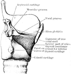
Dissection to show the lateral part of the cricothyroid membrane. The right ala of the thyroid cartilage has been removed.

Cricothyroid Membrane

Dissection to show the lateral part of the cricothyroid membrane. The right ala of the thyroid cartilage…
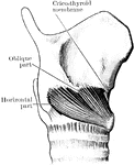
The cricothyroid muscle.

Cricothyroid Muscle

The cricothyroid muscle.

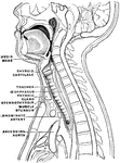
Diagram to show the course of the esophagus.

Course of the Esophagus

Diagram to show the course of the esophagus.

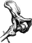
Obturator or thyroid dislocation of the hip.

Thyroid Dislocation of the Hip

Obturator or thyroid dislocation of the hip.

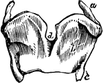
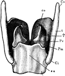
The more important cartilages of the larynx from behind. Labels: t, thyroid; Cs, its superior, and Ci, its inferior, horn of the right side; **, cricoid cartilage; †, arytenoid cartilage; Pv, the corner to which he posterior end of a vocal cord is attached; l'm, corner on which the muscles which approximate or separate the vocal cords are inserted; co, cartilage of Santorini.

Larynx

The more important cartilages of the larynx from behind. Labels: t, thyroid; Cs, its superior, and Ci,…

The larynx viewed from its pharyngeal opening. The back wall of the pharynx has been divided and its edges (11) turned aside. Labels: 1, body of hyoid; 2, its small, and 3, its great, horns; 4, upper and lower horns of thyroid cartilage; 5, mucous membrane of front of pharynx, covering the back of the cricoid cartilage; 6, upper end of gullet; 7, windpipe, lying in front of the gullet; 8, eminence caused by cartilage of Santorini; 9, eminence caused by cartilage of Wrisberg -- both lie in, 10, the aryteno-epiglottidean fold of mucous membrane, surrounding the opening (aditus laryngis) from pharynx to larynx; a, projecting tip of epiglottis; c, the glottis -- the lines leading from the letter point to the free vibrating edges of the vocal cords; b' the ventricles of the larynx -- their upper edges, marking them off from the eminences b, are the false vocal cords.

Larynx

The larynx viewed from its pharyngeal opening. The back wall of the pharynx has been divided and its…
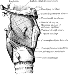
Dissection of the muscles in the lateral wall of the larynx. The right ala of the thyroid cartilage has been removed.

Larynx Muscle

Dissection of the muscles in the lateral wall of the larynx. The right ala of the thyroid cartilage…

A back view of the cartilages and ligaments of the larynx. Labels: 1, The posterior face of the epiglottis. 3, The os hyoides. 4, The lateral ligaments which connect the os hyoides and thyroid cartilage. 5, The posterior face of the thyroid cartilage. 6, The arytenoid cartilages. 7, The cricoid cartilage. 8, The junction of the cricoid and the arytenoid cartilages. 12, The first ring of the trachea.

A Back View of the Cartilages and Ligaments of the Larynx

A back view of the cartilages and ligaments of the larynx. Labels: 1, The posterior face of the epiglottis.…

A side view of the cartilages of the larynx. Labels: *, The front side of the thyroid cartilage. 1, The os hyoides (bone at the base of the tongue). 2, The ligament that connects the hyoid bone and thyroid cartilage. 3, 4, 5, The thyroid cartilage. 6, The cricoid cartilage. 7, The trachea.

A Side View of the Cartilages of the Larynx

A side view of the cartilages of the larynx. Labels: *, The front side of the thyroid cartilage. 1,…

Coronal section through larynx, to show its three compartments.

Coronal Section Through Larynx

Coronal section through larynx, to show its three compartments.

Mesial section through larynx, to show the outer wall of the right half.

Mesial Section Through Larynx

Mesial section through larynx, to show the outer wall of the right half.

Thyroid cartilages of the larynx.

Thyroid Cartilages of the Larynx

Thyroid cartilages of the larynx.

Front view of cartilages of larynx, trachea and Bronchi.

Front View of the Cartilages of the Larynx, Trachea and Bronchi.

Front view of cartilages of larynx, trachea and Bronchi.
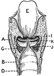
This illustration shows a longitudinal section of  the larynx as seen from behind (A. Thyroid Cartilage; B. Cricoid Cartilage; C. hyoid bone; D. Rings of trachea; E. Epiglottis; F. Arytenoid Cartilage; G. Thyro-arytenoid muscle; H. Adam's Apple; I. False Vocal Cords; J. True Vocal Cords; K. Ventricle; L. Rima Glottis).

Longitudinal Section of Larynx Seen from Behind

This illustration shows a longitudinal section of the larynx as seen from behind (A. Thyroid Cartilage;…
Sagittal section of a man.

Sagittal Section of the Neck and Head

Sagittal section of a man.

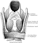
The lower part of the pharynx and the upper part of the esophagus have been slit up from behind, and the mucous membrane removed to show the muscular fibers. The two longitudinal bands are seen coming round to the front to be attached by a common tendon to the upper border of the cricoid cartilage.

Pharynx and Esophagus

The lower part of the pharynx and the upper part of the esophagus have been slit up from behind, and…

A side view of the thyroid and cricoid cartilages.

Thyroid

A side view of the thyroid and cricoid cartilages.

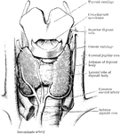
Dissection of the thyroid body and of the parts in immediate relation to it.

Thyroid Body

Dissection of the thyroid body and of the parts in immediate relation to it.

The trachea and bronchi. The thyroid body is indicated by a dotted line.

Trachea and Bronchi

The trachea and bronchi. The thyroid body is indicated by a dotted line.

Transverse section through the trachea and its immediate surroundings at the level of each of the upper five dorsal vertebrae.

Sections Through Trachea

Transverse section through the trachea and its immediate surroundings at the level of each of the upper…
"<em>A</em>, epiglottis; <em>B</em>, section of hyoid bone; <em>C</em>, false vocal cords which have nothing to do with the production of voice; <em>D</em>, oblong opening between the false and true vocal cords; <em>E</em>, true vocal cord;<em>F</em>, section of the thyroid cartilage; <em>H</em>, section of the anterior portion of the cricoid cartilage; <em>K</em>, trachea; <em>L</em>, section of the posterior portion of the cricoid cartilage; <em>M</em>, ladle-shaped cartilage." &mdash; Blaisedell, 1904

Vocal cords

"A, epiglottis; B, section of hyoid bone; C, false vocal cords which have…