Section through the pubic symphysis.

Cross Section of the Trunk Through the Pubic Symphysis

Section through the pubic symphysis.

Back view of the trunk.

Back View of the Bones of the Trunk

Back view of the trunk.

Diagram of horizontal section through upper part of 1st lumbar vertebra. The fine dots represent the great sac of the peritoneum, the coarse dots the lesser sac. Labels: A, aorta; Sp, spleen; B.D, bile duct; V.C, vena cava; P, pancreas; H.A, hepatic artery; K, kidney; L, liver; St, stomach; P.V, portal vein.

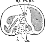
Horizontal Section Through Trunk

Diagram of horizontal section through upper part of 1st lumbar vertebra. The fine dots represent the…

Horizontal section through the trunk at the level of the first lumbar vertebra showing descriptive terms. Labels: M.P., mesial plane; S, sagittal; C, coronal or frontal.

Horizontal Section Through the Trunk

Horizontal section through the trunk at the level of the first lumbar vertebra showing descriptive terms.…

Frozen section across body at level of twelfth thoracic vertebra.

Section Across Trunk

Frozen section across body at level of twelfth thoracic vertebra.

Sagittal section through the trunk, 6 cm to the right of the median plane, viewed from the right. Note the close proximity of the duodenum and colon to the right kidney.

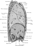
Side View of the Trunk

Sagittal section through the trunk, 6 cm to the right of the median plane, viewed from the right. Note…

The trunk showing rib, iliac crest, and pubis.

Side View of the Bones of the Trunk

The trunk showing rib, iliac crest, and pubis.

Transverse section through the middle of the first lumbar vertebra, showing the relations of the pancreas.

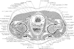
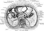
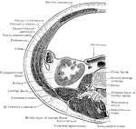
Transverse Section of the Trunk

Transverse section through the middle of the first lumbar vertebra, showing the relations of the pancreas.

Transverse section through the abdomen, opposite the second lumbar vertebra.

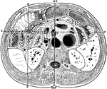
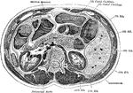
Transverse Section Through

Transverse section through the abdomen, opposite the second lumbar vertebra.

Diagram of vertical median section of abdomen. The fine dots represent the great sac of the peritoneum, the coarse dots the lesser sac. Labels: A, aorta; P, pancreas; I, intestine; R, rectum; L, liver; D, duodenum; B, bladder; St, stomach; c, colon; v, vagina.

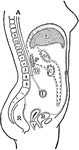
Vertical Median Section of the Trunk

Diagram of vertical median section of abdomen. The fine dots represent the great sac of the peritoneum,…

Showing the position of the viscera in the condition of visceroptosis (Glenard's disease). Labels: A, sterno-ensiform line: it crosses above the fifth costal cartilage; B, mid-epigastric line; C, umbilical line; D, mid-hypogastric line; a, pericardium; b, stomach (greatly elongated and dilated); c, liver; c', the lingual or Riedel's process; d, duodenum; e, caecum; f, transverse colon; g, rectum; h, elongated gastro-hepatic omentum.

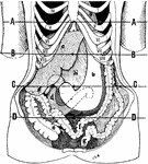
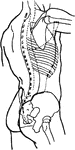
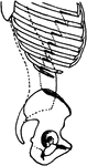
Position of the Viscera in the Condition of Visceroptosis

Showing the position of the viscera in the condition of visceroptosis (Glenard's disease). Labels: A,…