Clipart tagged: ‘villi’
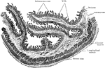
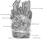
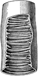
Longitudinal Section of Duodenum
Longitudinal section of duodenum; valvulae conniventes cut across, showing relation of these folds to…

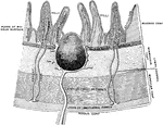
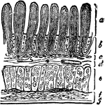
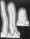
A Magnified Section of the Duodenum
A vertical section of the duodenum, highly magnified. Labels: 1, a fold-like villus; 2, epithelium,…
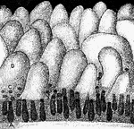
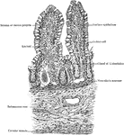
Mucous Membrane of the Ileum
The mucous membrane of the ileum. Labels: 1, cellular structure of the epithelium, or outer layer; 2,…
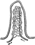
Intestinal Villus
An intestinal villus. They are little projections of the mucous membrane, covered with epithelium, and…
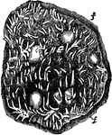
Section of Intestine wall
"A tiny block cut from the wall of the intestine showing villi and the mouths of glands at a; b, villus…
Transverse Section of Small Intestine
Transverse section of small intestine (lower part of duodenum), showing general arrangement of coats.
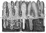
Mucous Membrane from the Jejunum
The mucous membrane from the jejunum. Labels: 1, Villi (folds of lining mucous membrane) in miniature.…
A Portion of the Mucous Membrane from the Small Intestine
Portion of the mucous membrane from the small intestine, magnified, showing the villi on its free surface,…
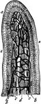
Rabbit's Intestinal Mucous Membrane
Vertical section of the intestinal mucous membrane of the rabbit. Two villi are represented, in one…
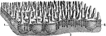
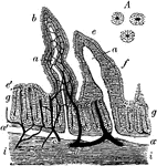
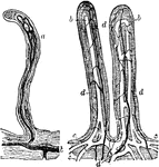
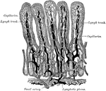
Small Intestine Villi
Villi of the small intestine, magnified about 80 diameters. In the left-hand figure the lacteals, a,b,c,…
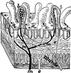
Glands and villi of the small intestine
"A, B, glands seen in vertical section with their orifices at C opening upon the membrane…
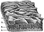
Piece of small intestine
"Piece of small intestine cut open to show wrinkling of inner coat bearing villi." —Davison, 1910
Villi of Small Intestine
Piece of the small intestine cut open to show wrinkling of inner coat bearing villi.
Transverse Section of Villi of Small Intestine
Transverse section of small intestine (jejunum), showing villi cut lengthwise.
Villi of the Small Intestine
Villi of the small intestine. Villa are minute, vascular processes which project from the mucous membrane…
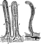
Villi of the Small Intestine
Villi of the small intestine; magnified about 80 diameters. In the right hand figure the lacteals, a,…
Villus of a Rat
Section of the villus of a rat killed during fat absorption. Labels: ep, epithelium; str, striated border;…
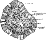
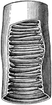
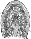
Transverse Section of Villus of Small Intestine
Transverse section of single intestinal villus, showing relation of epithelium, stroma, and vessels.