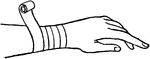
Clipart tagged: ‘wrist’
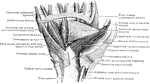
Muscles of the Arm
"Muscles on the front of the arm. Note the while cords, the tendons at the wrist." —Davison, 1910
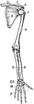
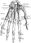
Bones of the Shoulder and Upper Extremity - Front View
"A, acromion; C, coracoid; CA, carpus; CL, clavicle; H, humerus; M, metacarpals; O, ventral surface…
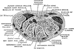
Transverse Section Through the Carpus
Transverse section through the carpus, showing the relative position of the tendons, vessels, and nerves.
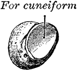
Cuneiform Bone
The cuneiform may be distinguished by its pyramidal shape, and by its having an oval, isolated facet…

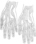
Hand and Bordure
"Argent, a sinister hand couped at the wrist and erected gules, within a bordure azure. BORDURE or BORDER.…
Dorsal Interosseous Muscles of the Hand
The dorsal interosseous muscles of the hand seen from the palmar aspect.
Palmar Interosseous Muscles of the Hand
The palmar interosseous muscles of the hand. Labels: P1, first; P2, second; and P3, third palmar interosseous…
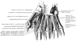
Tendon Sheaths of Wrist and Hand
Projections of two types of flexor tendon sheaths. Note that in the hand upon the right side there is…
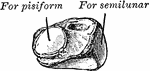
Pisiform Bone
The pisiform may be known by its small size and by its presenting a single articular facet. It is situated…
Carpal Articular Surface of the Radius
Carpal articular surface of the radius, and triangular fibro cartilage of the wrist.
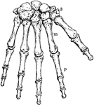
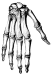
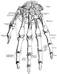
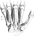
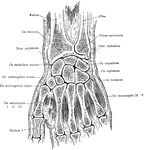
The Human Wrist and Hand Bones
Bones of the Wrist and Hand. Labels: m, metacarpal bones; p, phalanges; 3, bones of wrist.
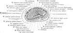
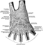
Cross Section Through the Wrist Joint and Carpal Bones
Section through the wrist joint and carpal bones.
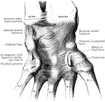
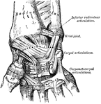
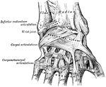
Ligaments of the Wrist
Ligaments on anterior aspect of radio carpal, carpal, and carpo metacarpal joints.
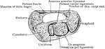
Transverse Section of Wrist
Transverse section through right wrist from above. The flexor tendons have been removed from the canal…