This human anatomy ClipArt gallery offers 96 illustrations of human coronary and pulmonary circulation of the cardiovascular system, which includes the organs and vessels involved in the flow of blood through the heart and lungs, where oxygen-depleted blood is sent to the lungs and oxygenated blood returns to the heart. Detailed views of the heart and views showing the relationship between the heart and lungs are also included in this category.
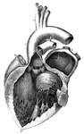
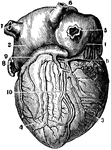
Human Heart
1 Right pulmonary veins; 1' Cavity of the auricle; 2 Wall of the auricle; 3,3' Walls of the ventricle;…
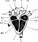
Interior of the Heart
Diagram of the interior of the heart. Labels: A, aorta; PA, pulmonary artery; VCI and VCS, vena cava…
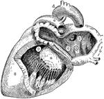
Left Side of Heart
Left side of heart. Labels: 1, cavity of left auricle (atrium); 3, opening of right pulmonary veins;…
Left Side of the Heart
Left side of the heart, showing the left auricle (atrium) and left ventricle).
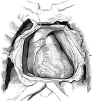
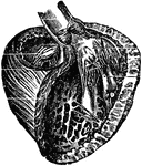
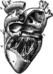
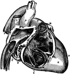
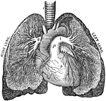
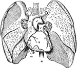
Posterior View of the Heart
A posterior view of the heart in a vertical position and with its vessels injected.
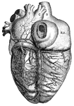
Right Side of Heart
Right side of heart. Labels: A, cavity of right ventricle; B, superior vena cava; C, inferior vena cava;…
Right Side of the Heart
Right side of the heart, showing the right auricle (atrium) and right ventricle).
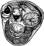
Section of Heart Showing Valves
Section of heart at level of valves. Labels: P, pulmonary artery, with flaps of semilunar valve open;…
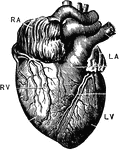
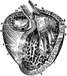
The Heart
The heart. Labels: RA, right auricle, RV; right ventricle; LA, left auricle; LV, left ventricle.
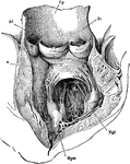
Ventricle of the Heart
The bases of the ventricle of the heart, showing the auriculoventricular, aortic, and pulmonary orifices…
The Pulmonary Artery
The pulmonary artery. Labels: t, The trachea. h, The heart. a, The aorta. p, The pulmonary artery. 1,…
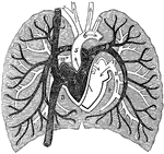
A Diagram of Pulmonary Circulation
A diagram of pulmonary circulation. Labels: 1, Descending vena cava. 2, Ascending cava vein. 3, Chamber…
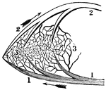
A Portion of the Pulmonic Circulation
A portion of the pulmonic circulation. 1, A branch of the artery that carries the impure blood to the…
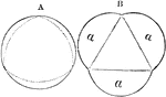
Action of Semilunar Valve
Section of the aorta, to show the action of the semilunar valve. A is intended to show the valves, represented…
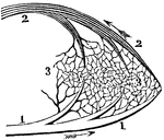
A Portion of the Systemic Circulation
A portion of the systemic circulation. 1, A branch of the aorta. This terminates in the capillaries…
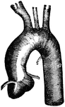
Thoracic Aorta
"The three branches from left to right are the unnamed ones. The primitive left carotid and the subclavian…
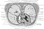
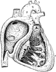
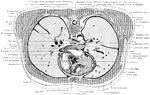
Cross Section of the Trunk Exposing the Ventricles of the Heart
Section exposing the ventricles of the heart.
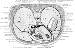
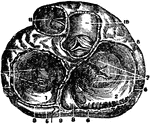
Cross Section of the Trunk Through the Inferior Portion of the Heart
Section through the inferior portion of the heart, exposing the dome of the diaphragm on the right side.
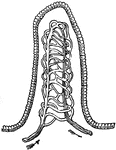
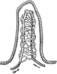
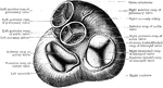
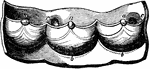
Semilunar Valves
Semilunar valves of the heart. The blood received by the left ventricle through the auriculo-ventricular…
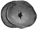
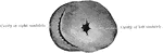
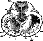
Ventricle
The right ventricle is in the shape of a crescent moon and the left ventricle is in the shape of a circle.
Ventricle and Aorta Laid Open
The left ventricle and the commencement of the aorta laid open. Mpm, Mpl, the papillary muscles. From…
Left Ventricle of the Heart
A three-quarter view of the left ventricle after removal of its anterior parietes.