This human anatomy ClipArt gallery offers 69 illustrations of human embryonic and fetal development, including external and dissected views of general development and development of specific systems progressing from fertilization to birth. Also includes views of the uterus and placenta as the embryo or fetus develops.

Section through the central canals of the spinal cord of a human embryo, showing ependymal (A) and neuroglial cells (B).

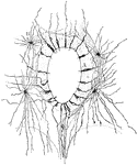
Section Through Spinal Cord Showing Neuroglial Cell

Section through the central canals of the spinal cord of a human embryo, showing ependymal (A) and neuroglial…

Three stages of development of the spinal cord. Labels: AC., anterior column; AH., anterior horn of gray matter; AL., alar lamina; AMF., antero-median fissure; AR., anterior nerve root; BC., column of Burdach; BL., basal lamina; E., ependyma; GC., column of Goll; LC., lateral column; MDL., mid-dorsal lamina; MVL., mid-ventral lamina, PC., early posterior column; PH., posterior horn of gray matter; PMF., postero-median fissure; PR., posterior nerve root.

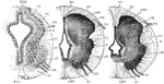
Development of Spinal Cord

Three stages of development of the spinal cord. Labels: AC., anterior column; AH., anterior horn of…

Diagram of development of spinal cord. Labels: c, central canal; af, anterior fissure; pf, posterior fissure; g, gray matter; w, white matter.

Development of the Spinal Cord

Diagram of development of spinal cord. Labels: c, central canal; af, anterior fissure; pf, posterior…

Diagram showing development of a spinal nerve and its components, together with the spinal sympathetic ganglia.

Development of Spinal Nerve

Diagram showing development of a spinal nerve and its components, together with the spinal sympathetic…

Development of the spinal nerves. A, Formation of nerve roots. B, Formation of nerve trunk (N).C, Formation of nerves. D, E, Formation of subordinate branches. F, Formation of nerve trunks in relation to the limb: dorsal and ventral trunks corresponding to lateral and anterior trunks in D and E. Labels: D.R., dorsal root; A1.C., alimentary canal; V.R., ventral root; Ao, aorta; N.T., neural tube; V., cardinal vein; No., notochord, M.P., muscle plate; D.G., dorsal ganglion; Sy., sympathetic cord; W.D., wolffian duct; Co., coelom; So., somatic division; V1, visceral branch; P, posterior primary division; Lat, lateral branches; Ant., anterior branches.

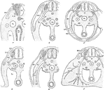
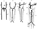
Development of the Spinal Nerve

Development of the spinal nerves. A, Formation of nerve roots. B, Formation of nerve trunk (N).C, Formation…

In the embryo the place of fibrous tissues is at first occupied by a mass of roundish cells, derived from the mesoblast. These develop into a network of branched cells or into groups of fusiform cells. Shown is a portion of submucous tissue of a uterus during pregnancy. Labels: a, branched cells, more or less spindel-shaped; b, bundles of connective tissue.

Tissue Growth of the Uterus

In the embryo the place of fibrous tissues is at first occupied by a mass of roundish cells, derived…

Diagrammatic view of a vertical transverse section of the uterus at the seventh week of pregnancy. Labels: c, c', cavity of uterus, which becomes the cavity of the decidua, opening at c, the cornua, into the Fallopian tubes, and at c' into the cavity of the cervix, which is closed by a plug of mucus; dv, decidua vera; dr, decidua reflexa, with the sparser villi imbedded in its substance; ds, decidua serotina, involving the more developed chorionic villi of the commencing placenta. The fetus is seen lying in the amniotic sac; passing up from the umbilicus is seen the umbilical pedicle of the yolk sac, which lies in the cavity between the amnion and chorion.

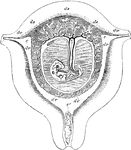
Uterus at Seventh Week of Pregnancy

Diagrammatic view of a vertical transverse section of the uterus at the seventh week of pregnancy. Labels:…

Section of the lining membrane of a human uterus at the period of commencing pregnancy showing the arrangement and other peculiarities of the glands, d, with their orfices, a, on the internal surface of the organ.

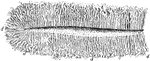
Lining Membrane of the Uterus

Section of the lining membrane of a human uterus at the period of commencing pregnancy showing the arrangement…

Diagram illustrating the development of the great veins. dc, ducts of Cuvier; j, jugular veins; h, hepatic vein; c, cardinal veins; s, subclavian vein; ci, inferior vena cava; r, renal veins; il, iliac veins; hij, hypogastric veins.

Development of Great Veins

Diagram illustrating the development of the great veins. dc, ducts of Cuvier; j, jugular veins; h, hepatic…