Vertical section of the ankle joint and foot. Labels: 1, tibia; 2, astragalus; 3, os calci; 4, scaphoides; 5, cuneiforme internum; 6, metatarsal bone of the great toe; 7, first phalanx of the great toe; 8, second phalanx of the great toe; 9, articular cavity between the tibia and astragalus; 10, synovial capsule between astragalas and calcis; 11, calcaneo-astragaloid interosseous ligament; 12, synovial capsule between astragalas and scaphoides; 13, calcaneo-scaphoid ligament; 14, calcaneo-cuboid ligament; 15, synovial capsule between scaphoides and cuneiforme internum; 16, synovial capsule between cuneiforme internum and first metatarsal bone; 17, metatarso-phalangeal articulation of the great toe, with sesamoid bones below; 18, phalangeal articulation of the great toe.

Ankle Joint and Foot

Vertical section of the ankle joint and foot. Labels: 1, tibia; 2, astragalus; 3, os calci; 4, scaphoides;…
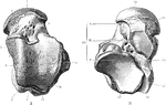
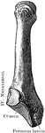
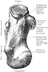
The right astragalus. A, Upper surface. B, Under surface. Labels: 1, groove for flex, long, hallucis; 2, internal tubercle; 3, trochlear surface for tibia; 4, body; 5, for articulation with internal malleolus; 6, head; 7, for articulation with internal malleolus; 8, neck; 9, for articulation with external malleolus; 10, surface against which the inferior tibio fibular ligament rests; 11, external tubercle; 12, external tubercle; 13, posterior, middle, and anterior facets for os calcis; 14, for articulation with navicular; 15, surface resting on inferior calcaneo-navicular ligament; 16, interosseous groove; 17, internal tubercle; 18, groove for the flexor longus hallucis.

Astragalus

The right astragalus. A, Upper surface. B, Under surface. Labels: 1, groove for flex, long, hallucis;…
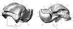
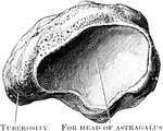
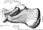
The right astragalus. A, As seen from the outer side. B, As seen from the inner side. Labels: 1, external tubercle; 2, groove for flexor longus hallucis; 3, internal tubercle; 4, surface against which the inferior tibio-fibular ligament rests; 5, trochlea for tibia; 6, for articulation with external malleolus; 7, neck; 8, head; 9, for articulation with navicular; 10, interosseous groove; 11, anterior, middle, and posterior facets for os calcis; 12, body; 13, surface resting on internal calcaneo navicular ligament; 14, for articulation with navicular; 15, head; 16, neck; 17, trochlea for tibia; 18, for articulation with internal malleolus; 19, body; 20, impression for internal lateral ligament; 21, internal tubercle; 22, groove for flexor longus hallucis; 23, external tubercle; 24, interosseous groove; 25, posterior and middle facets for os calis.

Astragalus

The right astragalus. A, As seen from the outer side. B, As seen from the inner side. Labels: 1, external…

The right cuboid bone (outer side).

Cuboid

The right cuboid bone (outer side).

The right cuboid bone (inner side).

Cuboid

The right cuboid bone (inner side).

Anterior view of the three cuneiform bones of the right foot.

Cuneiform Bones

Anterior view of the three cuneiform bones of the right foot.

The right external cuneiform (inner side).

External Cuneiform

The right external cuneiform (inner side).

The right external cuneiform (outer side).

External Cuneiform

The right external cuneiform (outer side).

The right internal cuneiform (inner side).

Internal Cuneiform

The right internal cuneiform (inner side).

The right internal cuneiform (outer side).

Internal Cuneiform

The right internal cuneiform (outer side).

The right middle cuneiform (inner side).

Middle Cuneiform

The right middle cuneiform (inner side).

The right middle cuneiform (outer side).

Middle Cuneiform

The right middle cuneiform (outer side).

The bones of the feet.

Bones of the Feet

The bones of the feet.

Bones of the foot, side view. In this figure the bones of the tarsus extend from the heel to <em>a</em>; the metatarsal bones are at <em>b</em>; and the bones of the toes are at <em>c</em>. In every movement of the foot there is some little motion between all these bones, and it is this that gives ease and grace to its motions.

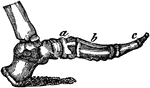
Side View of the Bone of the Foot

Bones of the foot, side view. In this figure the bones of the tarsus extend from the heel to a;…
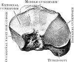
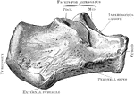
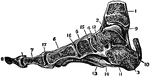
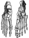
The bones of the foot. Labels: Ca, Calcaneum, or heel bone; Ta, articular surface for tibia on the astragalus; Cb, the cuboid bone

Bones of the Foot

The bones of the foot. Labels: Ca, Calcaneum, or heel bone; Ta, articular surface for tibia on the astragalus;…
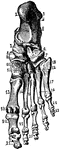
Bones of the foot (tarsus, metatarsus, and phalanges).

Bones of the Foot

Bones of the foot (tarsus, metatarsus, and phalanges).

Bones of the foot. At <em>e d f g h</em> are the 7 bones of the tarsus; at <em>a</em> are the 5 bones of the metatarsus; and at <em>b c</em> are the 14 bones of the toes (26 in all).

Bones of the Foot

Bones of the foot. At e d f g h are the 7 bones of the tarsus; at a are the 5 bones…
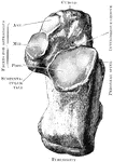
The bones of the foot. Labels: Ca, calcaneum, or os calcis; Ta, articular surface for tibia on the astragalus; N, scaphoid bone; CI, CII, first and second cuneiform bones; Cb, cuboid bone; M1, metatarsal bone of great toe.

Bones of the Foot

The bones of the foot. Labels: Ca, calcaneum, or os calcis; Ta, articular surface for tibia on the astragalus;…
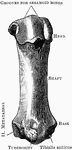
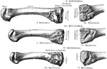
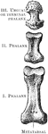
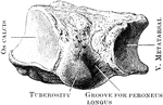
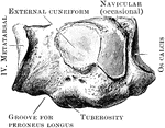
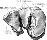
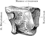
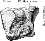
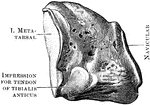
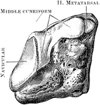
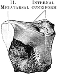
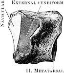
The first metatarsal bones of the right foot (plantar aspect).

First Metatarsal Bone

The first metatarsal bones of the right foot (plantar aspect).

View of the bases and shafts of the second, third, and fourth metatarsal bones of the right foot.

Metatarsal Bones

View of the bases and shafts of the second, third, and fourth metatarsal bones of the right foot.

Fifth right metatarsal bone (dorsal aspect).

Fifth Metatarsal

Fifth right metatarsal bone (dorsal aspect).

The right navicular bone as seen from behind.

Navicular Bone Viewed from Behind

The right navicular bone as seen from behind.

The right navicular bone viewed from the front.

Navicular Bone Viewed from the Front

The right navicular bone viewed from the front.

The right os calcis as seen from above.

Os Calcis Viewed from Above

The right os calcis as seen from above.

The right os calcis as seen from below.

Os Calcis Viewed from Below

The right os calcis as seen from below.

The os calcis as seen from the inner side.

Inner Side of Os Calcis

The os calcis as seen from the inner side.

The os calcis as seen from the outer side.

Outer Side of Os Calcis

The os calcis as seen from the outer side.

The phalanges of the toes (dorsal aspect).

Phalanges

The phalanges of the toes (dorsal aspect).