Horizontal preparation of cornea of frog; showing the network of branched cornea-corpuscles. The ground substance was completely colorless.

Magnified Frog Cornea Showing Branched Corpuscles

Horizontal preparation of cornea of frog; showing the network of branched cornea-corpuscles. The ground…

Surface view of part lamella of kitten's cornea, prepared first with caustic potash and then with nitrate of silver. By this method the branched cornea-corpuscles with their granular protoplasm and large oval nuclei are brought out.

Lamella of Kitten's Cornea

Surface view of part lamella of kitten's cornea, prepared first with caustic potash and then with nitrate…

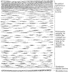
Vertical section of rabbit's cornea. Labels: anterior epithelium, showing the different shapes of the cells at various depths from the free surface; b, portion of the substance of cornea.

Magnified Rabbit's Cornea

Vertical section of rabbit's cornea. Labels: anterior epithelium, showing the different shapes of the…

Section of human cornea.

Section of Cornea

Section of human cornea.

Vertical section of rabbit's cornea, stained with gold chloride. Labels: e, Laminated anterior epithelium. Immediately beneath this is the anterior elastic lamina of Bowman. n, Nerves forming a delicate sub-epithelial plexus, and sending up fine twigs between the epithelial cells to end in a second plexus on the free surface; d, Descemet's membrane, consisting of a fine elastic layer, and a single layer of epithelial cells. the substance of the cornea, f, is seen to be fibrillated, and contains many layers of branched corpuscles, arranged parallel to the free surface, and here seen edgewise.

Section of Rabbit's Cornea

Vertical section of rabbit's cornea, stained with gold chloride. Labels: e, Laminated anterior epithelium.…
The cornea, which is the anterior transparent portion of the eyeball.

The Cornea

The cornea, which is the anterior transparent portion of the eyeball.

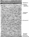
Vertical section of the cornea.

Vertical Section of the Cornea

Vertical section of the cornea.

A section of the human eye.

Eye

A section of the human eye.

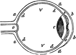
"a, sclerotic membrane; b, cornea; d, retina; o, optic nerve; v, vitreous humor." -Comstock 1850

Eye

"a, sclerotic membrane; b, cornea; d, retina; o, optic nerve; v, vitreous humor." -Comstock 1850
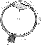
"Diagram of the eye. C., Cornea; a.h., aqueous humour; c.b., ciliary body; l., lens; I., iris; Sc., sclerotic; Ch., choroid; R., retina; v.h., vitreous humour; y.sp., yellow spot; n., optic nerve." -Thomson, 1916

Eye

"Diagram of the eye. C., Cornea; a.h., aqueous humour; c.b., ciliary body; l., lens; I., iris; Sc.,…
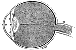
A section of the eye. Labels: 1, The sclerotic coat. 2, The cornea. 3, The choroid coat. 6, The iris. 7, The pupil. 8, The retina. 10, 11, Chambers or cavities of the eye that contain the aqueous humor. 12, The crystalline lens. 13, The vitreous humor. 15, The optic nerve. 14, 16, An artery of the eye.

A Section of the Eye

A section of the eye. Labels: 1, The sclerotic coat. 2, The cornea. 3, The choroid coat. 6, The iris.…

The eye of a common owl. The length from the anterior to posterior diameter lengthened.

Bird Eye

The eye of a common owl. The length from the anterior to posterior diameter lengthened.

"...and the cornea will become too flat, or not suffciently convex, to make the rays of light meet at the proper place, and the image will therefore tend to be formed beyond the retina, instead of before it..." -Comstock 1850

Cornea too Concave on Eye

"...and the cornea will become too flat, or not suffciently convex, to make the rays of light meet at…

"If the cornea is too convex, or prominent, the image will be formed before it reaches the retina, for the same reason, that of two lenses, that which is most convex will have the least focal distance." -Comstock 1850

Cornea too Convex on Eye

"If the cornea is too convex, or prominent, the image will be formed before it reaches the retina, for…
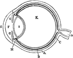
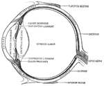
Plan of the eye seen in section. Labels: A, The Sclerotic Coat; B, The Choroid Coat; C, The Retina; D, The Optic Nerve; E, The Cornea; F, The Aqueous Humor; G, The Iris; H, The Ciliary Processes; I. The Crystalline Lens; K, The Vitreous Humor.

Diagram of the Eye

Plan of the eye seen in section. Labels: A, The Sclerotic Coat; B, The Choroid Coat; C, The Retina;…
The eye of a fish. The anterior to posterior diameter has been decreased.

Fish Eye

The eye of a fish. The anterior to posterior diameter has been decreased.

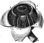
c, ciliary nerves going to be distributed in iris; d, smaller ciliary nerve; e, veins known as vasa vorticosa; h, ciliary ligament and muscle; k,l, converging fibres of iris; o, optic nerve.

Human Eye

c, ciliary nerves going to be distributed in iris; d, smaller ciliary nerve; e, veins known as vasa…
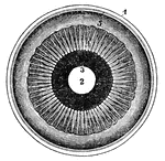
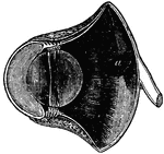
The iris and adjacent structures seen from behind. 1, the divided edge of the three coats, the choroid being the dark intermediate one; 2, the pupil; 3, the posterior surface of the iris; 4, the ciliary processes; 5, the scalloped anterior border of the retina.

Human Eye

The iris and adjacent structures seen from behind. 1, the divided edge of the three coats, the choroid…
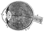
1, the sclerotic thicker behind than in front; 2, the cornea; 3, the choriod; 6, the iris; 7, the pupil; 8, the retina; 10, the anterior chamber of the eye; 11, the posterior chamber; 12, the crystalline lens, enclosed in its capsule; 13, the vitreous humour, enclosed in the hyaloid membrane and in cells formed in its interior by that membrae; 15, the sheath; and 16, the interior of the optic nerve, in the centre of which is a small artery.

Human Eye

1, the sclerotic thicker behind than in front; 2, the cornea; 3, the choriod; 6, the iris; 7, the pupil;…
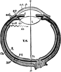
"Diagrammatic horizontal section of the eye of man. c, cornea; ch. choroid (dotted); C. P, ciliary processes; e. c, epithelium of cornea; e. cj, conjunctiva; f. c, yellow spot; I, iris; L, lens; ON, optic nerve; OS, ora serrata; o-x, optic axis; p. c. R, anterior non-visual portion of retina; P. E, pigmented epithelium (black); R, retina; sp. l, suspensory ligament; Scl, sclerotic; V. H, vitreous body." -Parker, 1900

Human Eye

"Diagrammatic horizontal section of the eye of man. c, cornea; ch. choroid (dotted); C. P, ciliary processes;…
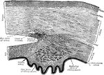
"Section of the human eye." — Blaisedell, 1904

Human eye

"Section of the human eye." — Blaisedell, 1904

Section of the eye, showing the relations of the cornea, sclera, and iris, together with the Ciliary muscle and the cavernous spaces near the angle of the anterior chamber.

Section of the Eye

Section of the eye, showing the relations of the cornea, sclera, and iris, together with the Ciliary…
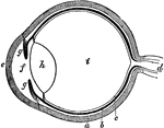
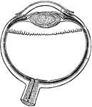
The eye. Labels: a, sclerotica; e, cornea; b, choroid; d, optic nerve; f, aqueous humor; g g , iris; h, lens; i, vitreous humor; c, retina.

The Eye

The eye. Labels: a, sclerotica; e, cornea; b, choroid; d, optic nerve; f, aqueous humor; g g , iris;…
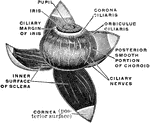
The middle or vascular coat of the eyeball exposed from without. Left eye, seen obliquely from above and before.

Vascular Coat of the Eye

The middle or vascular coat of the eyeball exposed from without. Left eye, seen obliquely from above…
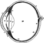
The left eyeball in horizontal section from before back. Labels: 1, sclerotic; 2, junction of sclerotic and cornea; 3, cornea; 4, 5, conjunctival membrane; 7, ciliary muscle; 10, choroid; 11, 13, ciliary processes; 14, iris; 15, retina; 16, optic nerve; 17, artery entering retina; 18, fovea centralis; 19, region where sensory part of retina ends; 26, 27, 28, are placed on the lens; 28 , suspensory ligament placed around lens; 29, vitreous humor; 30, aqueous humor in anterior chamber.

Left Eyeball in Horizontal Section

The left eyeball in horizontal section from before back. Labels: 1, sclerotic; 2, junction of sclerotic…
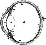
Diagram of a horizontal section through left eyeball and optic nerve.

Eyeball and Optic Nerve

Diagram of a horizontal section through left eyeball and optic nerve.

The left eyeball in horizontal section from before back. Labels: 1, sclerotic; 2, junction of sclerotic and cornea; 3, cornea; 4, 5, conjunctiva; 7, ciliary muscle; 10, choroid; 11, 13, ciliary processes; 14, iris; 15, retina; 16, optic nerve; 17, artery entering retina in optic nerve; 18, fovea centralis; 19, 20, region where sensory part of retina ends; 22, suspensory ligament; 24, the anterior part of the hyaloid membrane; 26, the lends; 29, vitreous humor; 30, aqueous humor.

The Eyeball in Horizontal Section

The left eyeball in horizontal section from before back. Labels: 1, sclerotic; 2, junction of sclerotic…
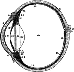
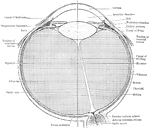
The left eyeball in horizontal section from before back. Labels: 1, sclerotic; 2, junction of sclerotic and cornea; 3, cornea; 4,5, conjunctiva; 6, posterior elastic layer of cornea; 7, ciliary muscle; 10, choroid; 11,13, ciliary processes; 14, iris; 15, retina; 16, optic nerve; 17, artery entering retina in optic nerve; 18, fovea centralis; 19, region where sensory part of retina ends; 22, suspensory ligament; 23 is placed in the canal of Petit, and the line from 25 points to it; 24, the anterior part of the hyaloid membrane; 26, 27, 28, are placed on the lends; 28 points to the line of attachment around it of the suspensory ligament; 29, vitreous humor; 30, anterior chamber of aqueous humor; 31, posterior chamber of aqueous humor.

Section of Left Eyeball

The left eyeball in horizontal section from before back. Labels: 1, sclerotic; 2, junction of sclerotic…
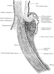
Section through ciliary region of eyeball.

Section Through Eyeball

Section through ciliary region of eyeball.

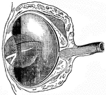
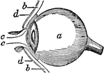
Side view of the eyeball. Labels: a, the eyeball, and b,b, are the upper and lower sides. Now in order to prevent dust and other bodies from working their way between the ball and the lid, through passages at c,c, into the socket of the eye where they would excite great inconvenience and pain, we find the common skin of the eye lids d,d, after covering their edges turn in a little way between the lid and the ball, and then turn back and are reflected over the surface of the cornea; where, to prevent the obstruction of vision, it becomes perfectly transparent.

Side View of the Eyeball

Side view of the eyeball. Labels: a, the eyeball, and b,b, are the upper and lower sides. Now in order…

"A representation of the manner in which the image is formed in the eye, when the cornea or crystalline lens is flattened. The perfect image would be produced at m m, behind the retina, and, of course, beyond the point necessary to secure perfect vision." —Wells, 1857

Flattened Eye

"A representation of the manner in which the image is formed in the eye, when the cornea or crystalline…
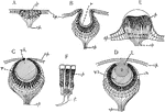
"Diagrams showing some of the stages in the increasing complexity of the simple eye in Invertebrates. A, simple pigment spot in epithelium having nerve-endings associated with pigment cells (as in some medusae); B, pigment cells in a pit-like depression (as in Patella); C, with pin-hole opening and vitreous humor in cavity (as in Trochus); D, completely closed pit, with lens and cornea (as in Triton and many other Mollusks); E, pigment area elevated instead of depressed, lens of thickened cuticula (as in the Medusa, Lizzia); F, retinal cells more highly magnified. ep., epidermis; f, nerve fibre; l, lens; op, optic nerve; p, pigment cells; r, retina; v.h., vitreous humor." -Galloway, 1915

Invertebrate Simple Eye

"Diagrams showing some of the stages in the increasing complexity of the simple eye in Invertebrates.…

The muscles of the eyeball, the view being taken from the outer side of the right orbit.

Eye Muscles

The muscles of the eyeball, the view being taken from the outer side of the right orbit.
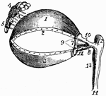
1, cartilage of the upper eyelid; 2, its lower border, showing the openings of the Meibomian glands; 3, the cartilage of the lower eyelids, also showing its border the openings of the Meibomian glands; 4, 5, the lachrymal glands; 6, its ducts; 7, the plica semilunaris; 8, the caruneula lachrymal canals; 10,11, the superior and inferior lachrymal canals; 12, the lachrymal sac; 13, the nasal duct, terminating at 14 in the lower meatus of the nose.

Eye Muscles

1, cartilage of the upper eyelid; 2, its lower border, showing the openings of the Meibomian glands;…

"A representation of the manner in which the image is formed in the eye of a near-sighted person. The curvature of the cornea, s s, and of the crystalline lens, c c, is so great that the image is formed at m m in advance of the retina." —Wells, 1857

Near-sighted

"A representation of the manner in which the image is formed in the eye of a near-sighted person. The…

"A representation of the manner in which the image is formed upon the retina in the perfect eye. The curvature of the cornea, s s, and of the crystalline lens, c c, is just sufficient to cause the rays of light proceeding from the image, e e, to converge the right focus, m, upon the retina." —Wells, 1857

Perfect Eye

"A representation of the manner in which the image is formed upon the retina in the perfect eye. The…
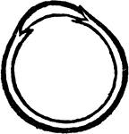
The retina of a human eye stretched out vertical.

Eye Retina

The retina of a human eye stretched out vertical.