Clipart tagged: ‘fibula’
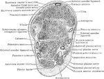
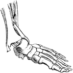
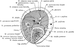
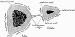
Section Through Ankle Joint
Coronal section through the left ankle joint, astragalus, and calcaneum.
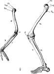
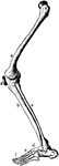
Arm and Leg Skeleton
The skeleton of the arm and leg. Labels: H, the humerus; Cd, its articular head which fits into the…

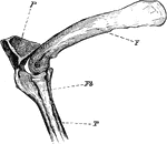
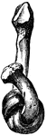
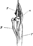
The Knee-joint of a Cormorant
"Phalacrocorax bicristatus. Cormorant. The knee-joint of a Cormorants. F, femur; P, patella; T, tibia;…
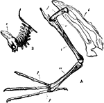
Diver Bones
"A. Pelvis and bones of the leg of the Leon or Diver; i, Innominate bone; f, Thighbone (femur); r, Tibia;…
Fibula
"A brooch, consisting of a pin, and of a curved portion furnished with a hook. The curved portion was…
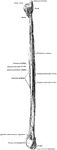
Fibula
The fibula (bone of leg). Labels: 1, head; 2, articular face; 3, insertion of external ligament; 4,…
Cross Section Five Inches Above the Lower End of the Fibula
Section five inches above the lower end of the fibula.
Cross Section Four Inches Above the Lower End of the Fibula
Section four inches above the lower end of the fibula.
Softened fibula
"The Fibula tied into a Knot after the Mineral Matter has been dissolved by Acid." — Blaisedell,…
Leg Bones of a Grebe
"F. Fibula; T, tibia, with a, its cnemial process, and P, large patella, of a grebe." Elliot Coues,…
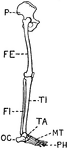
Human Leg (Front View)
This illustration shows a front view of a human leg. P. Pelvis, FE. Femur, TI. Tibia, FI. Fibula, TA.…
Human Leg (Front View), and Comparative Diagrams showing Modifications of the Leg
This illustration shows a human leg (front view), and comparative diagrams showing modifications of…
Human Leg (Side View)
This illustration shows a side view of a human leg. P. Pelvis, FE. Femur, TI. Tibia, FI. Fibula, TA.…
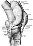
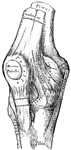
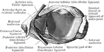
Knee Joint from Lateral Surface
Right knee joint from the lateral surface. The joint cavity and several bursae have been injected with…

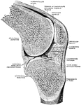
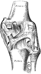
Sagittal Section Through Knee Joint
Right knee joint. Sagittal section through the external condyle of the femur. Mesal half of section,…
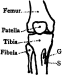
Bones of the Knee
Bones of the knee also showing muscle. Labels: s, insertion of the sartorius; g, insertion of the gracilis.
Leg Bones
"Bones of the leg. a, femur; b, tibia; c, fibula; d, tarsal bones; e, metatarsal bones; f, phalanges;…
Cross Section of Leg One Inch Above External Malleolus
Section one inch above the external malleolus.
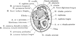
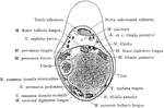
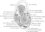
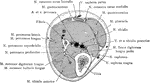
Cross Section Through Leg Three Inches Below Knee Joint
Section through the leg three inches below the right knee joint.
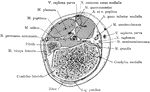
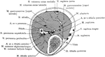
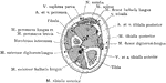
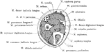
Cross Section of Leg, Two and a Half Inches above Ankle
Section two and a half inches above right ankle joint.
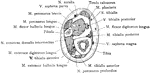
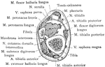
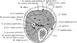
Cross Section Through Leg Two Inches Below Knee Joint
Section through the leg two inches below the right knee joint.
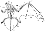
Noctule Bat
"Skeleton and volar Membranes of the Noctule Bat. c, clavicle; h, humerus; r, radius; u, ulna; d1, first…
Shank Bones
A section of the bones of the crus (shank of the leg) taken at about the middle of their length (schematized)…
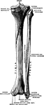
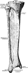
Tibia and Fibula
"The leg consists, like the forearm, of two bones. The larger, a strong, three-sided bone with…
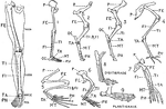
Vertebrate Appendages
"Diagrams of the girdles and appendages in a typical Vertebrate. A, anterior; B, posterior. ac., acetabulum,…
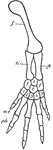
Vertebrate Hind Limb
"f., Femur; ti., tibia; fi., fibula; i., intermedium; t., tibiale (astragalus); f., fibulare (os calcis);…