Diagram showing the relation of the cerebrospinal to the sympathetic neurons. Labels: A, a medullated fiber, axon, or dendrite, coming from cerebrospinal system and dividing into numerous branches on reaching a sympathetic ganglion. These branches connect with those of the cells, b, B, in the ganglion, and these cells send their non-medullated fibers, axons, or dendrites, to supply the viscera, C, C, C, C.

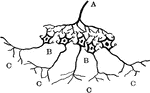
Diagram of the Relation between Cerebrospinal and Sympathetic Neurons

Diagram showing the relation of the cerebrospinal to the sympathetic neurons. Labels: A, a medullated…

Views of section of cervical cord. Labels: A, anterior surface; B, right side; C, upper surface; D, nerve roots; 1, anterior median fissure; 2, posterior median fissure; 3, anterior lateral depression; 4, posterior lateral groove; 5, anterior roots; 6, posterior roots; 6', ganglion; 7, spinal nerve; 7', posterior branch.

Sections of Cervical Spinal Cord

Views of section of cervical cord. Labels: A, anterior surface; B, right side; C, upper surface; D,…

The ninth, tenth, and eleventh cranial nerves. Labels: 1, Gasserian ganglion; 2, internal carotid; 3, pharyngeal branch of pneumogastric; 4, glosso-pharyngeal; 5, lingual; 6, spinal accessory; 7, middle constrictor of pharynx; 8, internal jugular vein; 9, superior laryngeal nerve; 10, ganglion of pneumogastric, 11, hypoglossal; 12, ditto communicating with eighth and first cervical; 13, external laryngeal; 14, second cervical; 15, pharyngeal plexus; 16, superior cervical ganglion; 17, superior cardiac nerve; 18, third cervical; 19, thyroid body; 20, fourth cervical; 21, recurrent laryngeal; 22, spinal accessory; 23, trachea 24, middle cervical ganglion; 25, middle cardiac nerve; 26, phrenic; 27, left carotid; 28, bronchial plexus; 29, phrenic; 30, inferior cervical ganglion; 31, pulmonary plexus; 32, aorta; 33, esophageal plexus; 34, vena axygos superior; 35, vena azygos minor; 36, gangliated cord of sympathetic.

The Ninths, Tenth, and Eleventh Cranial Nerves

The ninth, tenth, and eleventh cranial nerves. Labels: 1, Gasserian ganglion; 2, internal carotid; 3,…

"Dentalium: B, the shell of Dentalium Entalis, broken longitudinally, showing the animal in a contracted state. C, the shell, showing the animal advancing out; a and b, the foot, the lobes of which are developed in the form of a corolla; c, a part of the collar of the mantle. E, the animal, magnified, extricated from the shell, with them antle slit along the dorsal and medial line, detached in part from its posterior insertion, and turned aside so as to show the parts enclosed; a, the extremity of the foot, which closes the aperture j, of the collar lm, of the mantle nop; bb, lobes of the fot; c, the foot itself, presenting a depression or a channel, running its whole length; d, the head; e, the cerebral ganglion; f, f, the two sides of the mouth; g, g, the membranes which support the branchiae; gg, ii, the branchiae; pp, qq, the retractor muscles; s, the muscle of insertion; t, the expanded posterior extremity, in which is situated the vent." — Chambers' Encyclopedia, 1875

Dentalium

"Dentalium: B, the shell of Dentalium Entalis, broken longitudinally, showing the animal in a contracted…

Plan of the facial and intermediate nerves and their communication with other nerves.

Facial Nerves

Plan of the facial and intermediate nerves and their communication with other nerves.

An isolated ganglion cell of a human, showing sheath with nucleated cell lining, B. A Ganglion cell, with nucleus and nucleolus. C, Branched process. D. Unbranched process.

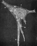
Ganglion Cell, Structure of

An isolated ganglion cell of a human, showing sheath with nucleated cell lining, B. A Ganglion cell,…

Different forms of ganglion cells. A, a, round ball-shaped unipolar cell from the human Gasserian ganglion. Two cells only show the process f; b, spindle shaped; c, multipolar ganglion cell from he spinal cord of the ox. d, D, Purkinjee ganglion cells from human cerebellum; ax, axis cylinder process; p, protoplasmic process; h, h, two cells surrounded with a nucleated sheath.

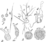
Different Forms of Ganglion Cells

Different forms of ganglion cells. A, a, round ball-shaped unipolar cell from the human Gasserian ganglion.…

Nerve cells, also known as nerve corpuscles, comprise the second principle element of the nervous tissue. The are not generally present in the nerve trunk, but are found in collections of nervous tissue called ganglia. Shown are the ganglion nerve corpuscles of different shapes.

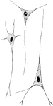
Ganglion Nerve Corpuscles

Nerve cells, also known as nerve corpuscles, comprise the second principle element of the nervous tissue.…
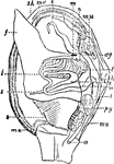
"Anatomy of an Acephalous Mollusc (Mactra): s, stomach; ii, intestine; ag, anterior ganglions; pg, posterior ganglions; mn, muscles; a, anus; h, heart; l, liver; f, foot; sh, shell; ma, mantle; b, branchiae; t, tentacula; r, oral, or respiratory syphon; t, anal syphon." — Chambers, 1881

Mollusc Anatomy

"Anatomy of an Acephalous Mollusc (Mactra): s, stomach; ii, intestine; ag, anterior ganglions; pg, posterior…
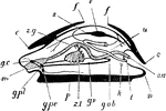
"Ideal mollusc. m., Mouth; g.c., cerebral ganglia; c., edges of mantle skirt; z.g., duct of right lobe of digestive gland; s., pericardial cavity; f., edges of shell-sac; v., ventricle of heart; u., nephridium; an., anus; n., posterior part of the foot; l., opening of nephridium; k., genital aperture; g.ab., abdominal ganglion on visceral loop; g.v., visceral ganglion; z.l., left lobe of digestive gland; p., foot; g.pe., pedal ganglion; g.pl. pleural ganglion." -Thomson, 1916

Mollusc Parts

"Ideal mollusc. m., Mouth; g.c., cerebral ganglia; c., edges of mantle skirt; z.g., duct of right lobe…

This illustration shows nerve cells from spinal ganglia.

Nerve Cells from Spinal Ganglia

This illustration shows nerve cells from spinal ganglia.

Reflex arc, as it is approximately in man. Labels: 1, nerve terminal, or sensory epithelium; 2, dendrite of sensory neuron; 3, cell body in dorsal root ganglion; 4, axon of sensory neuron; 5, dendrite of motor neuron; 6, cell body in ventral horn; 7, axon of motor neuron; 8, end organ (e.g., muscle-cell, gland-cell, etc.).

Nerve Reflex Arc

Reflex arc, as it is approximately in man. Labels: 1, nerve terminal, or sensory epithelium; 2, dendrite…

"<em>A</em>, branches of the nerves of smell; <em>B</em>, nerves of touch to the nostrils; <em>E, F, G,</em> nerves to the palate springing from a ganglion at <em>C</em>; <em>H</em>, a branch of the facial nerve, from which which other branches <em>D, I,</em> and <em>J</em>, spring to be distributed to the nostrils." — Blaisedell, 1904

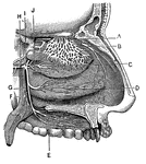
Nerves of the Nostril

"A, branches of the nerves of smell; B, nerves of touch to the nostrils; E, F,

Base of brain, spinal cord, and spinal nerves. Labels: V, 5th nerve; VI, 6th nerve; VII, a, facial nerve, b, auditory nerve; VIII, pneumo-gastric nerve; VIII, a, glosso-pharyngeal, b, spinal accessory; IX, hypoglossal; c1-c7, cervical nerve root; d1-d12, dorsal nerve roots; L1-L5, lumbar nerve roots; S4-S5, 4th and 5th sacral nerves; Cocc, coccygeal nerves; B.P., brachial plexus; L.P., lumbar plexus; S.P. sacral plexus; S a, b, c, cervical sympathetic ganglia.

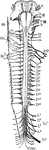
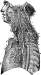
Spinal Cord, Spinal Nerves, and Base of Brain

Base of brain, spinal cord, and spinal nerves. Labels: V, 5th nerve; VI, 6th nerve; VII, a, facial nerve,…

Diagram showing anatomy of the spinal nerve roots and adjacent parts. Labels: G., gray matter of the spinal cord; W., white matter of the same; D.H., dorsal horn of gray matter; V.H., ventral horn of gray matter; D.R., dorsal root of spinal nerve; Sp.G., spinal ganglion; V.R., ventral root of the spinal nerve; Sp. N., spinal nerve; Rc., communicating branch (ramus communicans); S.G., sympathetic ganglion.

Spinal Nerve Roots

Diagram showing anatomy of the spinal nerve roots and adjacent parts. Labels: G., gray matter of the…
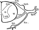
Diagram showing relation of neurons composing the spinal nerve roots with adjacent nervous structures. Labels: S.E., sensory epithelium connected by a sensory neuron with spinal cord; S.M., striated muscle receiving the axon from a motor-cell in the ventral horn of the gray matter in the cord; Sp. F., spinal fibers, medullated, sensory, and the motor, passing to the sympathetic ganglion where they connect with sympathetic neurons; S.F., S.F., non-medullated fibers from the sympathetic neurons passing to the viscera, the axons going to the plain muscle (P.M.), the dendrites to the sensory endings (S.E.).

Spinal Nerve Roots

Diagram showing relation of neurons composing the spinal nerve roots with adjacent nervous structures.…
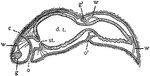
In this Turbellarian the digestive tract (d.t.) is a blind sac. st., boundary of stomodaeum and mesenteron; c, cilia; g, ganglion (brain); g', ganglion of a new individual which is being formed by fission; o, mouth; o', mouth of new individual in process of formation; w, excretory system.

Stenostoma

In this Turbellarian the digestive tract (d.t.) is a blind sac. st., boundary of stomodaeum and mesenteron;…

The sympathetic ganglions and their connection with other nerves. Labels: A, The semilunar ganglion and solar plexus. D, The thoracic (chest) ganglions. E, The external and internal branched of the thoracic ganglions. G, H, The right and left coronary (heart) plexus. I, N, Q , The inferior, middle and superior cervical (neck) ganglions. 1, The renal plexus of nerves. 2, The lumbar (loin) ganglions. 3, Their internal branches. 4, Their external branches. 5, The aortic plexus of nerves.

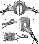
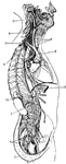
The Sympathetic Ganglions and their Connection to other Nerves

The sympathetic ganglions and their connection with other nerves. Labels: A, The semilunar ganglion…

General view of the sympathetic nervous. Labels: 1,2,3, cervical ganglia; 4, 1st thoracic ganglion; 5, 1st lumbar ganglion; 6, 7, sacral ganglion; 9, 9, cardiac nerves; 13, branch of pneumogastric nerve ending in semi-lunar ganglion; 14, epigastric plexus.

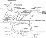
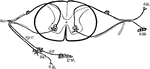
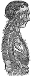
General View of the Sympathetic Nervous System

General view of the sympathetic nervous. Labels: 1,2,3, cervical ganglia; 4, 1st thoracic ganglion;…

"Diagrammatic section of spinal cord. p.f., Posterior fissure; p.c., posterior column of white matter; d.p.s., dorsal, posterior, sensory or afferent root; g., ganglion; v.a.m., ventral, anterior, motor or efferent root; c.n., compound spinal nerve with branches; s.g., sympathetic ganglion; a.c., anterior column--the anterior fissure is exaggerated; g.c., ganglion cells; g.m., grey matter; w.m., white matter." -Thomson, 1916

Vertebrate Spinal Cord

"Diagrammatic section of spinal cord. p.f., Posterior fissure; p.c., posterior column of white matter;…