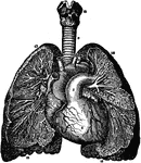
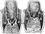
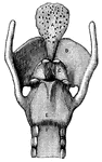
Branchi and Blood Vessels
Branchi of the lungs, the heart, and blood vessels. Labels: 1, left auricle; 2, right auricle; 3, left…
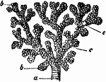
A Bronchial Tube
A small bronchial tube. Labels: a, dividing into its terminal branches, c; these have pouched or sacculated…
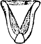
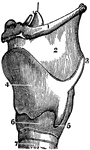
The Epiglottis
The epiglottis is a cartilaginous lid for the larynx. It is leaf-shaped, situated behind the base of…
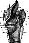
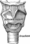
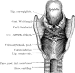
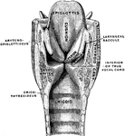
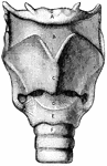
Larynx
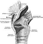
External view of the left side of Larynx. 1: Front portion of hyoid bone; 2: Upper edge of larynx; 3:…
Larynx
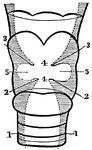
Cross section of the larynx above the vocal cords. 1: Right vocal cord. 2: Left vocal cord. 3: Cartilages…
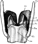
Larynx
The more important cartilages of the larynx from behind. Labels: t, thyroid; Cs, its superior, and Ci,…
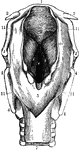
Larynx
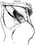
The larynx viewed from its pharyngeal opening. The back wall of the pharynx has been divided and its…
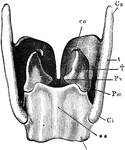
Larynx
The more important cartilages of the larynx from behind. Labels: t, thyroid; Cs, its superior, and Ci,…
Larynx
The larynx viewed from its pharyngeal opening. The back wall of the pharynx has been divided and its…
Larynx
The larynx is made of several pieces of gristle held together by muscle and other tissue. The largest…
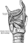
Side View of the Muscles of the Larynx
Muscles of the larynx. Side view. Right ala of thyroid cartilage removed.
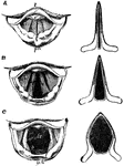
Larynx is Open and Shut Positions
Upper aperture of the larynx in the open (1) and shut (2) position. Labels: A, cushion of epiglottis;…
The Larynx Muscles of a Rook
"Muscles of the larynx. thyro-arytenoids, or openers of the glottis" Elliot Coues, 1884
The Larynx Muscles of a Rook
"Muscles of the larynx. Thyro-cricoids, posterior thyro-cricoids." Elliot Coues, 1884
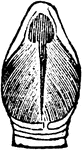
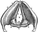
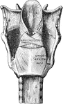
Larynx Seen from Behind
The larynx seen from behind after the removal of the muscles. The cartilages and ligaments only remain.
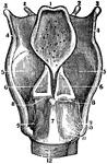
A View of the Larynx Showing the Vocal Ligaments
A view of the larynx showing the vocal ligaments. Labels: 1, The anterior edge of the larynx. 4, The…
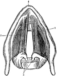
A Back View of the Cartilages and Ligaments of the Larynx
A back view of the cartilages and ligaments of the larynx. Labels: 1, The posterior face of the epiglottis.…
A Side View of the Cartilages of the Larynx
A side view of the cartilages of the larynx. Labels: *, The front side of the thyroid cartilage. 1,…

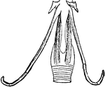
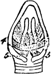
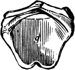
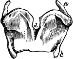
Arytenoid Cartilages of the Larynx
Arytenoid cartilages of the larynx. These are pitcher-like cartilages that are 2 in number, pyramidal-shaped,…
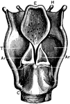
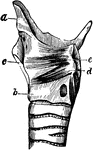
Back View of the Larynx
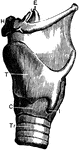
Labels: T, thyroid cartilage: C, cricoid cartilage; Tr, trachea; H, hyoid bone; E, epiglottis; I, joint…
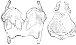
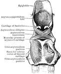
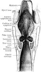
Cartilages from the Larynx
Cartilages from the larynx seen from the front. Labels: 1, vertical ridge of pomum Adami; 2, right ala;…
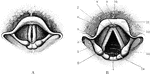
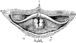
Cavity of Larynx
Cavity of larynx, as seen by means of the laryngoscope. A, the rima glottidis closed. B, the rima glottidis…
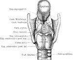
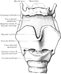
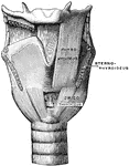
Front view of the larynx
"Cartilages and Ligaments of the Larynx. (Front view.) A, hyoid bone; B, membrane…
Front View of the Muscles of the Larynx
Muscles of the larynx, front view. The Sternothyroid and right Thyrohyoid have been removed.

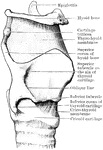
Lateral Aspect of Larynx
This illustration shows a lateral aspect of the larynx and its multiple parts (A. Thyroid Cartilage;…
Mesial Section Through Larynx
Mesial section through larynx, to show the outer wall of the right half.
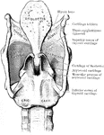
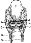
Posterior view of the larynx
"Cartilages and Ligaments of the Larynx. (Front view.) A, epiglottis; B, thyroid cartilage;…

The Larynx of a Rook
"Larynx viewed from before (below); a, thyroid bone or cartilage." Elliot Coues, 1884
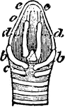
The Larynx of a Rook
"Larynx viewed from behind (above); a, thyroid bone; b, b, its appendages; c, cricoid; d, d, arytenoids;…
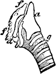
The Larynx of a Rook
"Larynx viewed from the right side; a, thyroid; b, appendage; c, cricoid; d, arytenoid; f, f, cartilage…
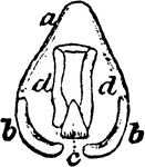
The Larynx of a Rook
"Larynx viewed from behind; a, thyroid; b, b, its appendages; c, cricoid; d, d, arytenoid." Elliot Coues,…
Section of the Larynx
A section of the larynx. Labels: 1, The trachea. 2, The lower vocal cords. 3, The upper vocal cords.…
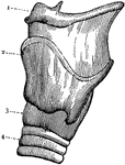
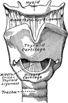
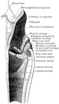
Side View of the Larynx
Labels: T, thyroid cartilage: C, cricoid cartilage; Tr, trachea; H, hyoid bone; E, epiglottis; I, joint…
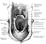
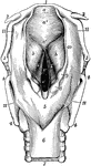
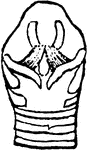
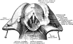
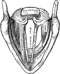
Superior Aperture of Larynx
Superior aperture of larynx, exposed by laying open the pharynx from behind.
The Larynx in Different Conditions of the Glottis
The larynx as seen by means of the laryngoscope in different conditions of the glottis. Labels: A, while…
Upper Part of the Larynx
View of the upper part of the larynx as seen by means of the laryngoscope during the utterance of a…
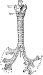
Front View of the Cartilages of the Larynx, Trachea and Bronchi.
Front view of cartilages of larynx, trachea and Bronchi.
Longitudinal Section of Larynx Seen from Behind
This illustration shows a longitudinal section of the larynx as seen from behind (A. Thyroid Cartilage;…
Lungs and Air Passages
The lungs and air passages seen from the front. On the left of the figure the pulmonary tissue has been…
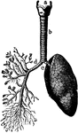
The Lungs and Air Passages
The lungs and air passages seen from the front. On the left of the figure the pulmonary tissue has been…
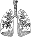
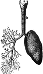
Lungs and Trachea
The lungs and windpipe (trachea). Labels: 1, larynx; 2, windpipe (trachea); 3, right lung, showing bronchi…