Diagram of muscle fiber with sarcolemma attached. Muscular tissue is the tissue by means of which the active movements of the body are produced. All muscular tissue consists of fibers, and whenever a muscle fiber contracts, it tends to bring its two ends, with whatever may be attached to them, together.

Muscle Fiber

Diagram of muscle fiber with sarcolemma attached. Muscular tissue is the tissue by means of which the…
Fiber-cells of plain muscular tissue, highly magnified.

Muscle Fiber Cells

Fiber-cells of plain muscular tissue, highly magnified.

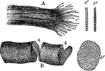
Fragments of striped (striated) muscle fibers, showing a cleavage in opposite directions, magnified 300 diameters. Striped muscle is nearly always under the control of the will, and is often spoken of as voluntary muscle. Labels: A, longitudinal cleavage; c, fibrillae separated from one another at the broken end of the fiber; c'c", single fibrils more highly magnified, in c; the elementary structures are square, in c" round; B, transverse cleavage; a, b, partially detached disks; b' detached disk, more highly magnified, showing the sarcous elements.

Fragments of Striped (Striated) Muscle Fibers

Fragments of striped (striated) muscle fibers, showing a cleavage in opposite directions, magnified…

Wave of contraction passing over a muscular fiber of dytiscus, very highly magnified. When a muscle contracts, the dark bands swell up and shorten (the light bands are also constricted), and the whole fiber broadens and shortens. Labels: R, R, portions of the fiber at rest; C, contracted part; I, I, intermediate condition.

Muscular Fiber Contracting

Wave of contraction passing over a muscular fiber of dytiscus, very highly magnified. When a muscle…

This illustration shows a diagram of nervous and cross-striate muscular tissue, showing the mode of connection between nerve fibres and muscle fibres. A, nerve cell (g) connected with muscle fibre (mf.) by nerve fibre (n.f.). The muscle fibre (m.f.) is composed of numerous fibrils (f) which are made up lengthwise of alternating discs of ligter and darker substance. These fibrils are shown more highly magnified in B and C. In B the fibril is uncontracted; in C it is contracted. D, nerve fibre more highly magnified showing a, axis; m, medullary sheath; and s, Schwann's sheath; ax., axon; d, dendron; n, node; n.m., nerve-muscle plate.

Muscular Tissue

This illustration shows a diagram of nervous and cross-striate muscular tissue, showing the mode of…
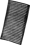
A fiber of cross-striped muscular tissue, showing the alternating bands.

Fiber of Muscular Tissue Showing Alternating Bands

A fiber of cross-striped muscular tissue, showing the alternating bands.

Fragment of a fiber of cross-striped muscular tissue, hardened, showing transverse cleavage.

Muscular Tissue Showing Transverse Cleavage

Fragment of a fiber of cross-striped muscular tissue, hardened, showing transverse cleavage.