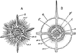
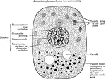
"Actinomma, a radiolarian with a shell and no mouth. A, whole animal with a portion of two spheres of shell removed. B, section, showing relation of protoplasm to the skeleton. c., central capsule; n, nucleus; p, protoplasm; o, openings through which the pseudopodia extend." -Galloway, 1915

Actinomma

"Actinomma, a radiolarian with a shell and no mouth. A, whole animal with a portion of two spheres of…
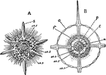
"Actinomma Asteracanthion, a Radiolarian with a limited number of specialized radii (axes), symmetrically arranged about the center. A, whole animal with portion of two spheres of shell removed. B, section, showing relation of the protoplasm to the skeleton. n, nucleus; p, protoplasm; sk., skeleton." — Galloway

Actinomma Asteracanthion

"Actinomma Asteracanthion, a Radiolarian with a limited number of specialized radii (axes), symmetrically…
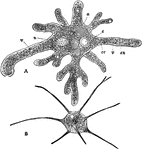
"Amaeba proteus; B, Amaeba radiosa. n, nucleus. c, contractile vacuole. a, food engulfed. ec, one of the nine pseudopods." —Davison, 1906

Amaeba Proteus

"Amaeba proteus; B, Amaeba radiosa. n, nucleus. c, contractile vacuole. a, food engulfed. ec, one of…
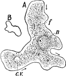
Amoung the simplest one-celled animals living in the ooze at the bottom of nearly every freshwater stream or pond is the Amoeba, whose body is barely visible to the unaided eye. Here we see the amoeba, highly magnified, showing(c.v.) pulsating vacuole; (f) food particle; (n) nucleus. (7B) The shape of the same individual 30 seconds later.

Amoeba

Amoung the simplest one-celled animals living in the ooze at the bottom of nearly every freshwater stream…
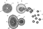
"Direct cell division (Amoeba). A, active specimen with pseudopodia; B, becoming spherical preliminary to division; C, beginning of elongation and constriction; D, later stage; E, daughter cells forming pseudopodia. ec, clear ectoplasm; en, granular endoplasm; f, food vacuole; n, nucleus; ps, pseudopodium; v, pulsating vacuole." -Galloway, 1915

Amoeba Cell Division

"Direct cell division (Amoeba). A, active specimen with pseudopodia; B, becoming spherical preliminary…
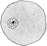
"In a general what we may describe a cell as a tiny mass of jelly in which floats another still smaller mass of slightly different composition, called the <em>nucleus</em> of the larger one."

Cell

"In a general what we may describe a cell as a tiny mass of jelly in which floats another still smaller…
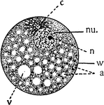
Diagram showing the principal parts of the cell and something of the protoplasmic architecture as it might appear while living. a, alveoli or spheres in the foam-work; c, centrosome; cy, cytoplasmic meshwork, containing granules; nu., nucleus; n, nucleolus; v. vacuole; w, cell wall.

Cell

Diagram showing the principal parts of the cell and something of the protoplasmic architecture as it…

Every human body begin as a single nucleated cell. This cell, known as the ovum, divides or segments and gives rise to a mass consisting of a number of similar units known as the morula. At this stage there are no distinguishable tissues.

Cell Development

Every human body begin as a single nucleated cell. This cell, known as the ovum, divides or segments…

Diagram showing the change which occur in the centrosomes and nucleus of a cell n the process of mitotic division (mitosis). The nucleus represented here has 4 chromosomes.

Cell Division

Diagram showing the change which occur in the centrosomes and nucleus of a cell n the process of mitotic…
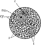
"Diagram showing the principal parts of the cell as it appeaers when killed and stained. The protoplasm shows more of a meshwork (cy), the spaces representing the alveoli. f, formed substance in alveoli." -Galloway, 1915

Cell Parts

"Diagram showing the principal parts of the cell as it appeaers when killed and stained. The protoplasm…
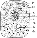
"Diagram of cell structure. Pl. Plastids in cytoplasm. cc. Centrosome. n. Nucleolus. Chr. Chromosomes. N. Nucleus. ct. General cytoplasm. V. Vacuole. Gr. Granules." -Thomson, 1916

Cell Structure

"Diagram of cell structure. Pl. Plastids in cytoplasm. cc. Centrosome. n. Nucleolus. Chr. Chromosomes.…

Diagram showing the structure of an animal cell.

Animal Cell

Diagram showing the structure of an animal cell.

Minute structure of a cell.

Cell, Diagram of

Minute structure of a cell.

A typical vegatable cell in a plant.

Vegetable Cell

A typical vegatable cell in a plant.

An illustration of the formation of the Cyclospora Cayetanensis egg-cell.

Formation of Cyclospora Cayetanensis Egg-cell

An illustration of the formation of the Cyclospora Cayetanensis egg-cell.
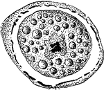
An illustration of the formation of the Cyclospora Cayetanensis egg-cell.

Formation of Cyclospora Cayetanensis Egg-cell

An illustration of the formation of the Cyclospora Cayetanensis egg-cell.
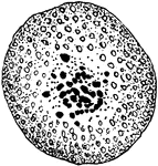
An illustration of the formation of the Cyclospora Cayetanensis egg-cell.

Formation of Cyclospora Cayetanensis Egg-cell

An illustration of the formation of the Cyclospora Cayetanensis egg-cell.

An illustration of the formation of the CCyclospora Cayetanensis spermatozooids

Formation of Cyclospora Cayetanensis Spermatozooids

An illustration of the formation of the CCyclospora Cayetanensis spermatozooids
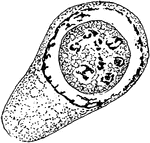
An illustration of the growth of a Cyclospora Cayetanensis Spore in the nucleus.

Growth of Cyclospora Cayetanensis Spore in Nucleus

An illustration of the growth of a Cyclospora Cayetanensis Spore in the nucleus.
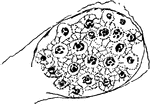
An illustration of the growth of spores of a Cyclospora Cayetanensis.

Formation of Cyclospora Cayetanensis Spores

An illustration of the growth of spores of a Cyclospora Cayetanensis.
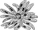
An illustration of the growth of spores of a Cyclospora Cayetanensis.

Formation of Cyclospora Cayetanensis Spores

An illustration of the growth of spores of a Cyclospora Cayetanensis.
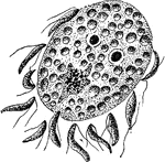
An illustration of the fertilization of the egg by the spermatozooids of a Cyclospora Cayetanensis.
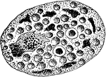
Fertilization of Cyclospora Cayetanensis

An illustration of the fertilization of the egg by the spermatozooids of a Cyclospora Cayetanensis.

An illustration of the fusion of egg and sperm-nuclei of a Cyclospora Cayetanensis.

Fusion of Egg and Sperm-nuclei of Cyclospora Cayetanensis

An illustration of the fusion of egg and sperm-nuclei of a Cyclospora Cayetanensis.
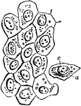
Flat epithelium cells from the surface of the peritoneum. Labels: a, cell body; c, nucleus.

Flat Epithelium Cells

Flat epithelium cells from the surface of the peritoneum. Labels: a, cell body; c, nucleus.

"Euglena viridis. A-D, four views illustrating euglenoid movements; E and H, enlarged views; F, anterior end further enlarged; G, resting form after binary fission; c. vac, contractile vacuole in H, reservoir in E and F; cy, cyst; fl, flagellum; m, mouth; nu, nucleus; oes, gullet; p, paramylum bodies; pg, pigment spot; r (in H), reservoir." -Parker, 1900

Euglena Viridis

"Euglena viridis. A-D, four views illustrating euglenoid movements; E and H, enlarged views; F, anterior…
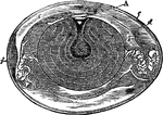
" Fig 110 - Hens egg, nat. size, in section; from Owen, after A. Thompson. A, cicatricle or "tread," with its nucleus, of white germ-yelk, floating on surface of pale thin nutritive yelk, leading to central yelk-cavity, x; a, the yellow yelk-ball, deposited in the successive layers, forming a set of halones, and enveloped in the chalaziferous membrane which is spun out at opposite poles into the twisted strings, chalazae, c, c; b, b', successive investments of softer white albumen; d, membrana putaminis, the "soft shell" or egg-pod, between layers of which at the great end of the egg is the air space, f;e, the shell." Elliot Coues, 1884

Hen's Egg

" Fig 110 - Hens egg, nat. size, in section; from Owen, after A. Thompson. A, cicatricle or "tread,"…

"One of the numberless microscopic bodies contained in semen, to which the seminal fluid owes its vitality, and which are the immediate and active means of impregnating or fertilizing the ovum of the female... I. broadest view; II. profile, of k, kernel or nucleus of the head, and m, filamentous body, ending in s, the long slender tail." —Whitney, 1889

Human Spermatozoa

"One of the numberless microscopic bodies contained in semen, to which the seminal fluid owes its vitality,…

"Nettling cells of Hydra. A, unexploded; B, exploded; b, barbs; c, the nettling cell in which the nettling organ is developed; cn., the cnidocil or "trigger"; cp., the capsule or nettling organ; f, the nettling filament or lasso; n, neck of the capsule; nu., nucleus of the cell." &mdash; Galloway

Hydra

"Nettling cells of Hydra. A, unexploded; B, exploded; b, barbs; c, the nettling cell in which the nettling…
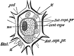
"Liteocircus annularis. cent. caps, central capsule; ext. caps. pr, extra-capsular protoplasm; int. cap. pr, intra-capsular protoplasm; nu, nucleus; psd, pseudopods; skel, skeleton; z, cells of zooxanthella." -Parker, 1900

L. Annularis

"Liteocircus annularis. cent. caps, central capsule; ext. caps. pr, extra-capsular protoplasm; int.…
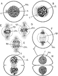
"Indirect or mitotic division; A, resting mother nucleus; B, coil stage, with the centrosomes separating; C, D (metaphase), and H, stages in the division of the chromosomes; F, diastroid (anaphase) stage; G and H show the return of the daughter nuclei to the coil and to the resting condition, and division of the cytoplasm, and the formation of the dividing wall: c, centrospheres; cl, chromatin coil; chr, chromosomes; nu., nucleus; n, nucleoius; sp, nuclear spindle; w, cell wall." &mdash; Galloway

Mitotic Division

"Indirect or mitotic division; A, resting mother nucleus; B, coil stage, with the centrosomes separating;…

"Plain muscle fibres. n, nucleus of muscle cell; p, undifferentiated cell protoplasm; p', the differentiated contractile portion of the cell." &mdash; Galloway

Muscle Fibres

"Plain muscle fibres. n, nucleus of muscle cell; p, undifferentiated cell protoplasm; p', the differentiated…

"Nematocyst of Hydra, showing cell-substance and nucleus, cyat, trigger hair, and everted thread." &mdash; Encyclopedia Britannica, 1893

Nematocyst

"Nematocyst of Hydra, showing cell-substance and nucleus, cyat, trigger hair, and everted thread." —…
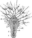
"A compound foraminiferan, Nodosaria. a, aperture of shell; f, food particles captured by the strands of protoplasm outside the shell; n, nucleus; sh, shell, 1-4, the successive chambers of the shell; 1, being the oldest." &mdash; Galloway

Nodosaria

"A compound foraminiferan, Nodosaria. a, aperture of shell; f, food particles captured by the strands…

The nucleus when in a condition of rest is bounded by a distinct membrane, possibly derived from the spongioplasm of the cell. Shown is a diagram of a resting nucleus.

Nucleus at Rest

The nucleus when in a condition of rest is bounded by a distinct membrane, possibly derived from the…

Diagrams of the nucleus showing the arrangement of chief chromatic filaments. A, Viewed from the side, the polar end being uppermost. Labels: p.c.f., primary chromatic filaments; n, nucleolus; n.o.m., node of meshwork. B, Viewed at the polar end. Labels: l.c.f., lopped chromatic filament; i.f., irregular filament.

Nucleus Showing Chromatic Filaments

Diagrams of the nucleus showing the arrangement of chief chromatic filaments. A, Viewed from the side,…
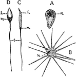
"Types of spermatozoa. A, from the round worm (Ascaris) with a cap, somewhat amaeboid; B, from the Crayfish, with numerous projections; C, from Frog; D, from Sea-urchin. h, head; m, middle piece; n, nucleus; t, tailor flagelium." &mdash; Galloway

Spermatozoa

"Types of spermatozoa. A, from the round worm (Ascaris) with a cap, somewhat amaeboid; B, from the Crayfish,…

"One of the numberless microscopic bodies contained in semen, to which the seminal fluid owes its vitality, and which are the immediate and active means of impregnating or fertilizing the ovum of the female... I. broadest view; II. profile, of k, kernel or nucleus of the head, and m, filamentous body, ending in s, the long slender tail." &mdash;Whitney, 1889
<p>Illustration of two spermatozoa from an ape.

Spermatozoa of an Ape

"One of the numberless microscopic bodies contained in semen, to which the seminal fluid owes its vitality,…
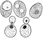
"Showing a Bit of Common Yeast Cake when mixed with Water and examined under the Microscope. There are large numbers of minute oval bodies, inside of which may commonly be seen one of more smaller bodies knows as <em>vacuoles</em>, shown in <em>a</em> and <em>b</em>. <em>c</em> shows a nucleus, <em>n</em>, inside of the yeast cell; <em>d shows a budding cell with the nucleus dividing; <em>e</em> shows the cell divided, the new cell containing a bit of the old nucleus." &mdash; Blaisedell, 1904

Yeast cake

"Showing a Bit of Common Yeast Cake when mixed with Water and examined under the Microscope. There are…