Clipart tagged: ‘thorax’
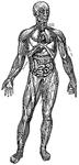
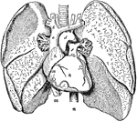
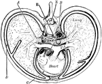
Principal Organs of the Thorax and Abdomen
"The principal muscles are seen on the left, and superficial veins on the right." — Blaisedell, 1904
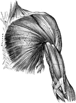
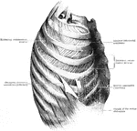
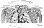
Muscles of the Arm and Thorax
Muscles of the thorax and front of the arm, showing some of the boundaries of the axilla.
Natural and Contracted Chest
The left figure represents the natural shape of the chest, and that upon the right, the contracted state…
Diaphragm
View of the diaphragm; 1, cavity of the thorax; 2, diaphragm separating the cavity of the thorax from…
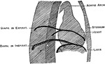
Diaphragm During Expiration
Diaphragm in the state of its greatest ascent in expiration; 2, muscles of the abdomen in action forcing…
Diaphragm During Inspiration
Diaphragm in its state of greatest descent in inspiration; 2, muscles of the abdomen, showing the extend…
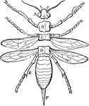
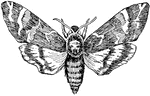
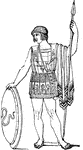
Parts of an Insect
"A, the head; B, C, D, segments of the thorax; E, abdomen; F, ovipositor." -Cooper, 1887
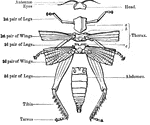
External Anatomy of an Insect Skeleton
"Anatomy of the external skeleton of an insect" — Goodrich, 1859
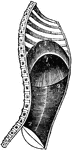
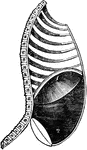
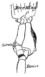
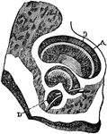
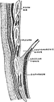
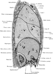
Pleural Sac
Left pleural sac in a subject hardened by formalin injection, opened into by the removal of the costal…
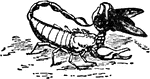
Scorpion
"Drawing from tlife of the Italian scorpion Euscorpius italicus, Herbst, holding a blue-bottle fly with…
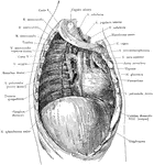
Thoracic Cavity after Removal of Lung
Deep structures of the right thoracic cavity, after removal of the right lung.
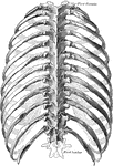
Thorax
"The ribs are long, flat, and curved bones which bend round the chest somewhat like the hoops…
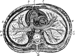
Thorax
"The transverse section of the thorax. 1, anterior mediastinum; 2, internal mammary vessels; 3, triangularis…
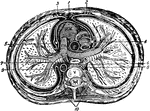
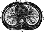
Thorax
"Cross-section of thorax. A, bronchus, entering the lung; B, the aorta cut at its origin and again at…
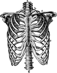
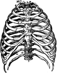
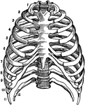
Thorax
The skeleton of the thorax. Labels: a, g, vertebral column; b, first rib; c, clavicle; d, third rib;…
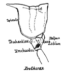
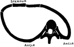
The Thorax
The thorax. Labels: a, the sternum. b to c, the true ribs; d to h, the false ribs; g, h, the floating…
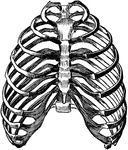
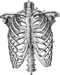
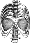
Human Thorax (Chest)
Thorax. The thorax, or chest, is an elongated conical-shaped cage, formed by the sternum and costal…

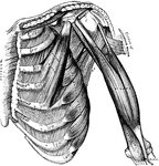
Thorax and Abdomen
"Thorax and abdomen. 1, 1, 1, 1. Muscles of the chest. 2, 2, 2, 2. Ribs. 3, 3, 3. Upper, middle and…
Side View of the Thorax and Part of the Abdomen
Lateral, sagittal section through the left thorax and upper portion of abdomen, viewed from the left.…
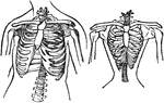
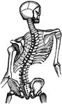
Changes in the Thorax following Scoliosis of the Spine
Showing the changes in the thorax which follow scoliosis of the spine. The convexity of the spinal curvature…
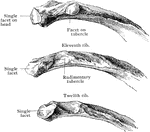
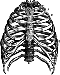
Thorax Skeleton
The skeleton of the thorax. Labels: a, g, vertebral column; b, first rib; c, clavicle; e, seventh rib;…
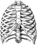
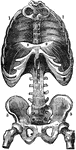
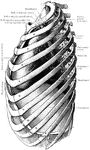
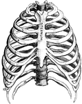
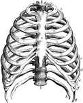
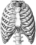
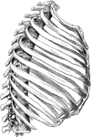
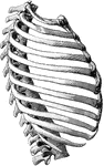
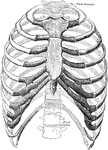
The Bones of the Thorax
Front view of the bones of the thorax, including the ribs, sternum and vertebrae. Labels: 1, first bone…
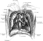
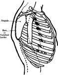
Dissection of the Thorax
Topography of the retrocardiac structures of the mediastinum, after the removal of the heart and pericardium.
Frontal Section Through the Thorax
Frontal section through the thorax, passing through the mediastinum and the middle of the humeral heads.
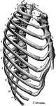
Lower Half of the Thorax
The lower half of the thorax, with four lumbar vertebrae showing the diaphragm from above. Labels: 1,2,3,…
Orthodiagram of the Thorax
Orthodiagram of the thorax. The position of parts is shown in extreme inspiration; the position of the…
The Thorax
Diagram of a transverse section of the thorax. Labels: 1, anterior mediastinum; 2, internal mammary…
Transverse Section of the Thorax
Transverse section of the thorax. Labels: 1, anterior mediastinum; 2, internal mammary vessels; 3, triangularis…
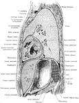
Side View of the Trunk
Sagittal section through the trunk, 6 cm to the right of the median plane, viewed from the right. Note…