Diagrammatic transverse section of abdomen, to show the peritoneum on transverse tracing. A, at level of foramen of Winslow. B, lower down. In A note, one of the vasa brevia arteris passing to the stomach between the layers of the gastrosplenic omentum, and also the foramen of Winslow leading into the lesser sac which lies behind the stomach.

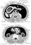
Transverse Section of Abdomen

Diagrammatic transverse section of abdomen, to show the peritoneum on transverse tracing. A, at level…

The right auricle (atrium) and ventricle of the heart opened, and a part of their right and anterior walls removed, so as to show their interior; 1/2, 1, superior vena cava; 2, inferior vena cava; 2', hepatic veins cut short; 3, right auricle; 3', placed in the fossa ovalis, below which is the Eustachian valve; 3", is placed close to the aperture of the coronary vein; +, +, placed n the auriculo-ventricular groove, where a narrow portion of the adjacent walls of the auricle and ventricle has been preserved; 4, 4, cavity of the right ventricle; the upper figure is immediately below the semilunar valves; 4', large columna carnea or musculus papillaris; 5, 5', 5", tricuspid valve; 6, placed in the interior o the pulmonary artery, a part of the anterior wall of that vessel having been removed, and a narrow portion of it preserved at its commencement where the semilunar valves are attached; 7, concavity of the aortic arch close to the cord of the ductus arteriosus; 8, ascending part of sinus of the arch covered at its commencement by the auricular appendix and pulmonary artery; 9, placed between the innominate and left carotid arteries; 10, appendix of the left auricle; 11, 11, the outside of the left ventricle, the lower figure near the apex.

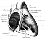
Right Atrium and Ventricle of the Heart

The right auricle (atrium) and ventricle of the heart opened, and a part of their right and anterior…

Branchi of the lungs, the heart, and blood vessels. Labels: 1, left auricle; 2, right auricle; 3, left ventricle; 5, pulmonary artery; 6, arch of the aorta; 7, superior vena cava; 8, arteria innominata; 9, left primitive carotid artery; 10, left subclavian artery; 11, trachea; 12, larynx; 13, upper lobe of right lung; 14, upper lobe of left lung; 15, trunk of right pulmonary artery; 16, lower lobes of the lungs.

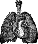
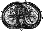
Branchi and Blood Vessels

Branchi of the lungs, the heart, and blood vessels. Labels: 1, left auricle; 2, right auricle; 3, left…
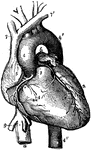
Front view of the heart and great vessels. The pulmonary artery has been cut short close to its origin. Labels: 1, right ventricle; 2, left ventricle; 3, root of the pulmonary artery; 4, 4', arch of the aorta; 4'', the descending thoracic aorta; 5, part of the right auricle; 6, part of the left auricle; 7, 7', innominate veins joining to form the vena cava superior; 8, inferior vena cava; 9, one of the large hepatic veins; X, placed in the right auriculo-ventricular groove, points to the right or posterior coronary artery; X, X, placed in the anterior interventricular groove, indicate the left or anterior coronary artery.

Heart

Front view of the heart and great vessels. The pulmonary artery has been cut short close to its origin.…

Diagram of the heart completely divided into right and left halves, and of a double (systematic and pulmonary) circulation. Ad, right auricie; Vcs, anterior vena cava; Vd, right ventricle; Ap, pulmonary artery; P, lung; Vp, pulmonary vein; As, left auricle; Vs, left ventricle; Ao, aorta; D, gut; L, liver; Vp, portal vein; Lv, hepatic vein.

Heart Divided into Left and Right Halves

Diagram of the heart completely divided into right and left halves, and of a double (systematic and…

The cavities of the right auricle and right ventricle of the heart.

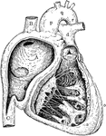
Auricle and Ventricle of the Heart

The cavities of the right auricle and right ventricle of the heart.

The right auricle and ventricle opened, and a part of their right and anterior walls removed, so as to show their interior. Labels: 1, superior vena cava; 2, inferior vena cava; 2', heptic veins cut short; 3, right auricle; 3', placed in the fossa ovalis, below which is the Eustachian valve; 3", is placed close to the aperture of the coronary adjacent walls of the auricle and ventricle has been preserved; 4, cavity of the right carnea or musculus papillaris; 5, 5', 5", tricuspid valve; 6, placed in the interior of the pulmonary artery, a part of the anterior wall of that vessel having been removed, and a narrow portion of it preserved at its commencement, where the semilunar valves are attached; 7, concavity of the aortic arch close to the cord of the ductus arteriosus; 8, ascending part or sinus of the arch covered at its commencement by the auricular arteries; 10, appendix of the left auricle; 11, the outside of the left ventricle, the lower figure near the apex.

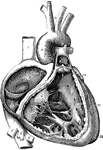
A Diagram of the Heart

The right auricle and ventricle opened, and a part of their right and anterior walls removed, so as…

Diagram of the interior of the heart. Labels: A, aorta; PA, pulmonary artery; VCI and VCS, vena cava inferior and vena cava superior; T, tricuspid, and M, mitral valve. The other letters same as in preceding figure.

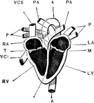
Interior of the Heart

Diagram of the interior of the heart. Labels: A, aorta; PA, pulmonary artery; VCI and VCS, vena cava…

Right side of heart. Labels: A, cavity of right ventricle; B, superior vena cava; C, inferior vena cava; a, wall of right ventricle; b, c, columnae carneae; d, pulmonary vein; e, f, tricuspid valve; m, semilunar valve; o, wall of left ventricle; p, q, v, ascending aorta, arch and descending aorta.

Right Side of Heart

Right side of heart. Labels: A, cavity of right ventricle; B, superior vena cava; C, inferior vena cava;…

Diagram of a transverse section of the thorax. Labels: 1, anterior mediastinum; 2, internal mammary vessels; 3, triangularis sterni muscle; 4, right phrenic nerve between pleura and pericardium; 5, left phrenic nerve between the pleura and pericardium; 6, thoracic duct in posterior mediastrinum; 7, esophagus with left vagus in front and right vagus behind; 8, vena azygos major; 9, thoracic aorta giving off intercostal arteries; 10, gangliated cord of sympathetic; R.V., right ventricle; R.A., right auricle of heart in middle mediastinum; P.A., pulmonary artery; A., aorta; C., vena cava superior; V. dorsal vertebra.

The Thorax

Diagram of a transverse section of the thorax. Labels: 1, anterior mediastinum; 2, internal mammary…

Transverse section of the thorax. Labels: 1, anterior mediastinum; 2, internal mammary vessels; 3, triangularis sterni; 4, 5, phrenic nerves; 6, thoracic duct; 7, esophagus; 8, vena azygos major; 9, thoracic aorta; 10, sympathetic; R. V., right ventricle; R. A., right auricle; P. A. pulmonary artery; A., aorta; C., superior vena cava; V., dorsal vertebra.

Transverse Section of the Thorax

Transverse section of the thorax. Labels: 1, anterior mediastinum; 2, internal mammary vessels; 3, triangularis…

Sketch of the principal venous trunks. Labels: 1, superior vena cava; , inferior vena cava; 3, right subclavian and innominate veins; 4, left subclavian and innominate veins; 5, 5, right and left internal jugular veins; 8, right azygos vein; 10, left azygos vein; 13, 13, common iliac veins; 14, 14, sacral veins.

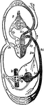
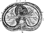
Principal Venous Trunks

Sketch of the principal venous trunks. Labels: 1, superior vena cava; , inferior vena cava; 3, right…