Vertical section of area pellucida and area opaca (left extremity of figure) of blastoderm of a fresh laid egg (unincubated). Labels: S, superficial layer corresponding to epiblast; D, deeper layer, corresponding to hypoblast; and probably in part to mesoblast; M, large "formative cells," filled with yolk granules, and lying on the floor of the segmentation cavity; A, the white yolk immediately underlying the segmentation cavity.

Blastoderm of an Egg

Vertical section of area pellucida and area opaca (left extremity of figure) of blastoderm of a fresh…
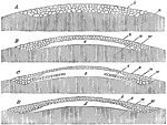
"Further development of hen's egg; after Haeckel: A, the mulberry mass of cleavage cells, b, same as seen on top in fig 111, F, here viewed in profile in section, resting upon n, the simply-shaded part of the figure, to represent conventionally the mass of food-yelk. A, morula stage (as before); B, blastula stage, the mass of cells, b, forming the blastoderm, uplifted from the food-yelk, leaving the cleavage-cavity, s; w, the thickened rim of the germ-disc; C, the blastula in process of inversion, by which a layer of entoderm-cells, i, growing from periphery to centre, will apply itself to the layer of exoderm-cells, e, obliterating the cleavage-cavity, s; D, the disc-gastrula completed, by union of entoderm, i, with exoderm, e, leaving the primitive intestinal cavity, d, which is quite similar in appearance to the cleavage cavity, s, but morphologically quite different." Elliot Coues, 1884

Egg Germination

"Further development of hen's egg; after Haeckel: A, the mulberry mass of cleavage cells, b, same as…
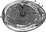
" Fig 110 - Hens egg, nat. size, in section; from Owen, after A. Thompson. A, cicatricle or "tread," with its nucleus, of white germ-yelk, floating on surface of pale thin nutritive yelk, leading to central yelk-cavity, x; a, the yellow yelk-ball, deposited in the successive layers, forming a set of halones, and enveloped in the chalaziferous membrane which is spun out at opposite poles into the twisted strings, chalazae, c, c; b, b', successive investments of softer white albumen; d, membrana putaminis, the "soft shell" or egg-pod, between layers of which at the great end of the egg is the air space, f;e, the shell." Elliot Coues, 1884

Hen's Egg

" Fig 110 - Hens egg, nat. size, in section; from Owen, after A. Thompson. A, cicatricle or "tread,"…
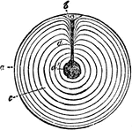
"Meroblastic ovum (yelk) of domestic fowl, bat. size, in section; after haeckel. a, the thin yelk-skin, enclosing the yellowfood-yelk, which is deposited in concentric layers, c, d; b, the cicatricle or tread with its nuclues, whence passes a cord of white yelk (here represented in black) to the central cavity, d'" Elliot Coues, 1884

Fowl Ovum

"Meroblastic ovum (yelk) of domestic fowl, bat. size, in section; after haeckel. a, the thin yelk-skin,…
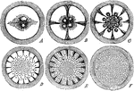
"The first change in the parent-cell is that by which it becomes broken up into a mass of cells, each of which is just like itself. This process is called segmentation of the vitellus; each one of the numerous resulting cells is called a cleavage-cell. The nucleus of the parent-cell divides into two; each attracts its half of the yelk; the halves furrow apart and there are now two cleavage cells in place of the one parent-cell a furrow at right angles to the first, and redivision of the nuclei; results in four cleavage-cells. Radiating furrows intermediate to the first two bisect the four cells, and would render eight cells, were not these simultaneously doubled by a circular furrow which cleaves each, with the result of sixteen cleavage-cells. So the subdivision goes on until the parent-cell becomes a mass of cells. This particular kind of cleavage, by radiating and concentric furrowing, is called discoidal, and the resulting heap of little cells assumes the figure of a thin, flat, circular disc. Segmentation of the vitellus, in whatever manner it may go on, results in a mulberry-like mass of cleavage-cells; and the original cytula has become what is called a morula. This process is shown closely here." Elliot Coues, 1884

The Segmentation of the Vitellus

"The first change in the parent-cell is that by which it becomes broken up into a mass of cells, each…
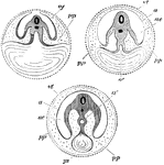
Diagram showing the three successive stages of development. Transverse vertical sections. The yolk sac, ys, is seen progressively diminishing in size. In the embryo itself the medullary canal and notochord are seen in section. a', in middle figure, the alimentary canal, becoming pinched off, as it were, from the yolk sac; a', in right hand figure, alimentary canal completely closed; a, in last two figures amnion; ac', cavity of amnion filled with amniotic fluid; p, space between amnion and chorion continuous with the pleuroperitoneal cavity inside the body; vt, vitelline membrane; ys, yolk sac, or umbilical vesicle.

Development of the Yolk Sac

Diagram showing the three successive stages of development. Transverse vertical sections. The yolk sac,…

Diagram of the various stages of cleavage of the yolk.

Stages of Cleavage of the Yolk

Diagram of the various stages of cleavage of the yolk.