The Human Sensory Systems: Sight ClipArt gallery offers 189 illustrations related to human vision.
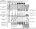
Plan of the retinal neurons.

Plan of the Retinal Neurons

Plan of the retinal neurons.

Diagram of the structure of the human retina. Labels: I, pigment layer; II, rod and cone layer; R, rods; C, cones; III-IX, intraretinal nerve-elements; X, axons which pass to optic nerve.

Retinal Structure

Diagram of the structure of the human retina. Labels: I, pigment layer; II, rod and cone layer; R, rods;…
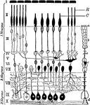
The rods of Corti of the ear showing a pair of rods separated from the rest. Labels: i, inner, and e, outer rods.

The Rods of Corti

The rods of Corti of the ear showing a pair of rods separated from the rest. Labels: i, inner, and e,…

The rods of Corti of the ear. Shown hear is a bit of the basilar membrane with several rods on it, showing how they cover in the tunnel of Corti. Labels: i, inner, and e, outer rods;, b, basilar membrane; r, reticular membrane.

The Rods of Corti

The rods of Corti of the ear. Shown hear is a bit of the basilar membrane with several rods on it, showing…

A. a shortsighted eye; B. an arrow which it attempts to perceive, but is prevented by the convergence of the passage of the visual rays to foci, at C., before they reach the retina at D. E. the same eye similarly situated, showing how by the interventions of a concave lens, G., the rays are diverged, and the image of the arrow, F., accurately converged to the retina at A.

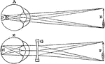
Shortsighted Vision and Correction

A. a shortsighted eye; B. an arrow which it attempts to perceive, but is prevented by the convergence…
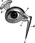
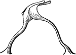
The tear gland of the eye. The tears are carried from this factory b little ducts (b) and are poured over the surface of the eye. They serve to keep the eye moist, so that it can be moved about in its socket easily by the muscles. They also serve to wash out substances that get into the eye. The tears go into openings (c c) and pass through two ducts which unite in one (d e) which ends in the nose.

Tear Gland

The tear gland of the eye. The tears are carried from this factory b little ducts (b) and are poured…

An illustration of a transverse section of an ideal eye. "A, summit of cornea; SC, sclerotic; S, Schlemm's canal; CH, choroid; I, iris; M, ciliary muscle; R, retina; N, optic nerve; HA, aqueous humour; L, crystalline lens, the anterior of the double lines on its face showing its form during accommodation; HV, vitreous humour; DN, internal rectus muscle YY', principle optical axis; C, [center] of the ocular globe..." (Britannica, 132).

Transverse Section of an Ideal Eye

An illustration of a transverse section of an ideal eye. "A, summit of cornea; SC, sclerotic; S, Schlemm's…
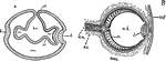
"Diagrams illustrating two stages in the development of the vertebrate eye. A, showing the relation of the ectoderm, the brain vesicle, and the optic vesicle. The right side of the figure shows a later stage than the left. B, later stage, showing the lens, eye-ball and retina in position. b.v., brain vesicle formed by the invagination oof the ectoderm (ect.); l, lens; mes., mesodermal tissue; o.n., optic nerve; o.s., optic stalk; o.v., optic vesicle, a portion of the brain vesicle; r, retinal layer; v.h., interior of eye-ball which comes to contain the vitreous humor." — Galloway

Vertebrate Eye

"Diagrams illustrating two stages in the development of the vertebrate eye. A, showing the relation…
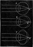
Diagram illustrating rays of light converging in a normal eye (A), a myopic eye (B), and a hypermetropic eye (C)

Vision

Diagram illustrating rays of light converging in a normal eye (A), a myopic eye (B), and a hypermetropic…