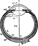
"Diagrammatic horizontal section of the eye of man. c, cornea; ch. choroid (dotted); C. P, ciliary processes; e. c, epithelium of cornea; e. cj, conjunctiva; f. c, yellow spot; I, iris; L, lens; ON, optic nerve; OS, ora serrata; o-x, optic axis; p. c. R, anterior non-visual portion of retina; P. E, pigmented epithelium (black); R, retina; sp. l, suspensory ligament; Scl, sclerotic; V. H, vitreous body." -Parker, 1900

Human Eye

"Diagrammatic horizontal section of the eye of man. c, cornea; ch. choroid (dotted); C. P, ciliary processes;…
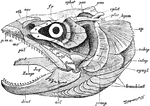
"Salmo fario, the entire skull, from the left side. art, articular; branchiost, branchiostegal rays; dent, dentary; epiot, epiotic; eth, supra-ethnoid; fr, frontal; hyom, hyomandibular; intop, inter-opercular; pal, palantine; par, parietal; pmx, pre-maxilla; praop, pre-opercular; pt, pterygoid; pter, pterotic; Quad, quadrate; socc, supra-occipital; sphot, sphenotic; subop, sub-opercular; sympl, symplectic; Zunge, basi-hyal." -Parker, 1900

Fish Skull

"Salmo fario, the entire skull, from the left side. art, articular; branchiost, branchiostegal rays;…
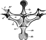
"Rana esculenta. The shoulder girdle from the ventral aspect. Co, coracoid; Co', epicoracoid; Cl, clavicle; G, glenoid cavity; Ep, episternum; Fe, fenestra between procoracoid and coracoid; KC, cartilage separating scapula and clavicle; Kn, xiphisternum; m, junction of epicoracoids; S, scapula; St, sternum." -Parker, 1900

Edible Frog Shoulder Girdle

"Rana esculenta. The shoulder girdle from the ventral aspect. Co, coracoid; Co', epicoracoid; Cl, clavicle;…

"Rana esculenta. Pelvic girdle from the right side. G, acetabulum; Il, P, ilium; Is, ischium; Kn, pubis." -Parker, 1900

Edible Frog Pelvic Girdle

"Rana esculenta. Pelvic girdle from the right side. G, acetabulum; Il, P, ilium; Is, ischium; Kn, pubis."…

"Skeleton of crocodile. C, causal region; D, thoracic region of spinal column; F, fibula; Fe, femur; H, humerus; I, ischium; L, lumbar region; R, radius; Ri, ribs; S, sacrum; Sc, scapula; Sta, abdominal ribs; T, tibia; U, ulna." -Parker, 1900

Crocodile Skeleton

"Skeleton of crocodile. C, causal region; D, thoracic region of spinal column; F, fibula; Fe, femur;…
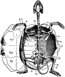
"Cistudo lutaria. Skeleton seen from below; the plastron has been removed and is represented on one side. C, costal plate; Co, coracoid; e, entoplastron; Ep, epiplastron; F, fibula; Fe, femur; H, humerus; Il, ilium; Is, ischium; M, marginal plates; Nu, nuchal plate; Pb, pubis; Pro, pro-coracoid; Py, pygal plates; R, radius; Sc, scapula; T, tibia; U, ulna." -Parker, 1900

Marsh Turtle Skeleton

"Cistudo lutaria. Skeleton seen from below; the plastron has been removed and is represented on one…

"Chelone midas. Transverse section of skeleton. C, costal plate; C', centrum; M, marginal plate; P, lateral element of plastron; R, rib; V, expanded neural plate." -Parker, 1900

Green Turtle Skeleton

"Chelone midas. Transverse section of skeleton. C, costal plate; C', centrum; M, marginal plate; P,…
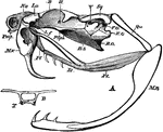
"A, lateral view of skull of rattlesnake (Crotalus). B. O, basi-occipital; B. S, basi-sphenoid; E. O, exoccipital; F. O, fossa ovalis; La, conjoined lacrymal and pre-fontal; L. f, articulation between lacrymal and frontal; Mn, mandible; Mx, maxilla; Na, nasal; Pl, palatine; Pmx, pre-maxilla; P. Sph, pre-sphenoid; Pt, pterygoid; Qu, quadrate; Sq, squamosal; II. V, foramina of exit of the second and fifth cranial nerves. B, transverse section at point lettered B in Fig. .A; T, trabeculae." -Parker, 1900

Rattlesnake Skull

"A, lateral view of skull of rattlesnake (Crotalus). B. O, basi-occipital; B. S, basi-sphenoid; E. O,…
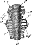
"Columba livia. Sacrum of a nestling (about fourteen days old), ventral aspect. c1, centrum of first sacral vertebra; c1, centrum of fifth caudal; c. r, first sacral rib; l1, centrum of first lumbar; l3, of third lumbar; s1, of fourth lumbar; s3, of sixth lumbar; tr. p, transverse process of first lumbar; tr. p', of fifth lumbar; tr. p'', of first sacral." -Parker, 1900

Rock Pigeon Sacrum

"Columba livia. Sacrum of a nestling (about fourteen days old), ventral aspect. c1, centrum of first…
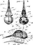
"Columba livia. Skull of young specimen. A, dorsal; B, ventral; C, left side. al. s, alisphenoid; au, angular; ar, articular; b. o, basi-occipital; d, dentary; e. o, ex-occipital; eu, aperture of Eustachian tube; f. m, foramen magnum; fr, frontal; i.o. s, inter-orbital septum; ju, jugal; lc, lachrymal; lb. s, lambdoidal suture; m. eth, mesethmoid; mx, maxilla; mx. p, maxillo-palatine process; na, na', na'', nasal; o. c, occipital condyle; or. fr, orbital plate of frontal; pa, parietal; pa. s, parasphenoid (rostrum); pl, palatine; p. mx, premaxilla; pt, pterygoid; qu, quadrate; s. an, supra-angular; s. o, supra-occipital; sq, squamosal; ty, tympanic cavity; IL-XII, foramina for cerebral nerves." -Parker, 1900

Rock Pigeon Skull

"Columba livia. Skull of young specimen. A, dorsal; B, ventral; C, left side. al. s, alisphenoid; au,…

"Columba livia. Left manus of a nestling. The cartilaginous parts are dotted. cp. 1, radiale; cp. 2, ulnare; mcp. 1, 2, 3, metacarpals; ph. 1, phalanx of first digit; ph. 2, ph. 2', phalanges of second digit; ph. 3, phalanx of third digit; ra, radius; ul, ulna." -Parker, 1900

Rock Pigeon Manus

"Columba livia. Left manus of a nestling. The cartilaginous parts are dotted. cp. 1, radiale; cp. 2,…

"Columba livia. Left innominate of a nestling. The cartilage is dotted. ac, acetabulum; a. tr, anti-trochanter; il, pre-acetabular; and il', post-acetabular portion of ilium; is, ischium; i s. f, ischiatic foramen; ob. f, obturator notch; pu, pubis." -Parker, 1900

Rock Pigeon Innominate

"Columba livia. Left innominate of a nestling. The cartilage is dotted. ac, acetabulum; a. tr, anti-trochanter;…

"Columba livia. Part of left foot of an unhatched embryo. The cartilage is dotted. mtl. 2, second; mtl. 3, third; and mtl. 4, fourth metatarsal; ti, tibia; tl. 1, proximal tarsal cartilage; tl. 2, distal tarsal cartilage." -Parker, 1900

Rock Pigeon Embryo Foot

"Columba livia. Part of left foot of an unhatched embryo. The cartilage is dotted. mtl. 2, second; mtl.…
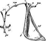
"Lepus cuniculus. Shoulder-girdle with anterior end of sternum of young specimen. a, acromion; af, pre-scapular fossa; c, coracoid; cl, ossified clavicle; ma, meta-cromion; mss, meso-scapular segment; ost, pre-sternum; pc, pre-coracoid; pf, post-scapular fossa; sr, sternal ribs." -Parker, 1900

Rabbit Shoulder Girdle

"Lepus cuniculus. Shoulder-girdle with anterior end of sternum of young specimen. a, acromion; af, pre-scapular…

"Nearly perfect fossil skeleton of Archaegosaurus." -Taylor, 1904

Archegosaurus

"Nearly perfect fossil skeleton of Archaegosaurus." -Taylor, 1904

The skeleton of Ichthyosaurus.

Ichthyosaurus

The skeleton of Ichthyosaurus.

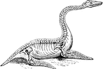
The skeleton of Plesiosaurus.

Plesiosaurus

The skeleton of Plesiosaurus.

The skull of the pterodactyle.

Pterodactyle Skull

The skull of the pterodactyle.

The wing skeleton of the pterodactyle.

Pterodactyle Wing

The wing skeleton of the pterodactyle.

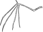
The skeleton of a bat's wing.

Bat Wing

The skeleton of a bat's wing.

"Fossil skull of the mososaurus." -Taylor, 1904

Mososaurus Skull

"Fossil skull of the mososaurus." -Taylor, 1904

"The mammoth (Elephas primigenius)." -Taylor, 1904

Mammoth

"The mammoth (Elephas primigenius)." -Taylor, 1904

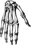
Bones of the hand.

Bones of the Hand

Bones of the hand.

Bones of the forearm.

Bones of the Forearm

Bones of the forearm.

Section through the bones and ligaments of the foot. The parts of the joints are well shown.

Bones and Ligaments of the Foot

Section through the bones and ligaments of the foot. The parts of the joints are well shown.

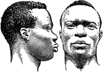
in early development of race, the Negro type were found in Africa.

Early Races, Negro

in early development of race, the Negro type were found in Africa.

in early development of race, the Mongolian type consistent of Kalmucks, Chinese, and Amerindians.

Early Races, Mongolian

in early development of race, the Mongolian type consistent of Kalmucks, Chinese, and Amerindians.

in early development of race, the Caucasian type consisted of Mediterranean men (Jews of Algiers), Mediterranean woman (Berber) and Nordics (Englishmen).

Early Races, Caucasian

in early development of race, the Caucasian type consisted of Mediterranean men (Jews of Algiers), Mediterranean…

Early developments of racial types, a tomb paint from an Egyptian tomb.

Racial Types From Egyptian Paintings

Early developments of racial types, a tomb paint from an Egyptian tomb.

Perhaps the earliest people to form real cities in the western region of the world, were a people of mysterious origin called Sumerians. This drawing is a very early Sumerian stone carving showing Sumerian warriors in phalanx.

Stone Carvings of Sumerian Warriors

Perhaps the earliest people to form real cities in the western region of the world, were a people of…
An early figure of the Egyptian hippopotamus goddess.

Egyptian Hippopotamus Goddess

An early figure of the Egyptian hippopotamus goddess.

Bones of the head.

Bones of the Head

Bones of the head.

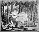
Singular warfare of the American Indians. Caption below illustration: "I no longer hesitated; I took my aim; discharged ,y piece; and the animal was instantly stretched before me with a groan, which I conceived to be that of a human creature. I went up to it, and judge my astonishment when I found I had killed an Indian."

American Indians Warfare

Singular warfare of the American Indians. Caption below illustration: "I no longer hesitated; I took…
Men carrying someone in a palanquin in China.

Palanquin

Men carrying someone in a palanquin in China.

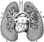
"The Lungs. 1, Summit of lungs. 2, Base of lungs. 3, Trachea. 4, Right bronchus. 5, Left bronchus. 6, Left aurical of heart. 7, Left superior pulmonary vein. 8, Right superior pulmonary vein. 9, Left ventricle of heart. 10, Right ventricle of heart." -Foster, 1921

Lungs

"The Lungs. 1, Summit of lungs. 2, Base of lungs. 3, Trachea. 4, Right bronchus. 5, Left bronchus. 6,…
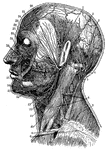
"Superficial arteries and nerves of the face and neck. 1, Temporal artery; 2, artery behind the ear; 3, occipital artery; 4, greater occipital nerve; 5, smaller occipital nerve; 6, nerve of the neck; 7, trapezius muscle; 8, clavicular nerves; 9, clavicle; 10, sterno-cleido-mastoid muscle; 11, outer artery of the head; 12, inner artery of the head; 13, salivary gland; 14, nerves of the lower jaw; 15, outer maxillary artery; 16, nerve of the chin; 17, circular muscle of the mouth; 18, greater yoke muscle; 19, nerves below the eye; 20, masseter, or chewing muscle; 21, ear passage; 22, arteries of the forehead; 23; nerves of the forehead; 24, eye-closing muscle; 25, facial artery; 26, facial nerve." -Foster, 1921

Arteries and Nerves

"Superficial arteries and nerves of the face and neck. 1, Temporal artery; 2, artery behind the ear;…

"Nerves of the had. 1, Nerves of the skin; 2, tendons; 3, arteries of the palm of the hand; 4, elbow nerve; 5, elbow artery; 6, nerve of the forearm; 7, nerve of the under-arm; 8, artery of the under-arm." -Foster, 1921

Hand Nerves

"Nerves of the had. 1, Nerves of the skin; 2, tendons; 3, arteries of the palm of the hand; 4, elbow…
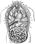
"Thorax and abdomen. 1, 1, 1, 1. Muscles of the chest. 2, 2, 2, 2. Ribs. 3, 3, 3. Upper, middle and lower lobes of the right lung. 4, 4. Lobes of the left lung. 5, Right ventricle of the heart. 6. Left ventricle. 7. Right auricle. 8. Left auricle. 9. Pulmonary artery. 10. Aorta. 11. Descending vena cava. 12. Trachea. 13. Oesophagus. 14, 14, 14, 14. Pleura. 15, 15. Diaphragm. 16, 16. Right and left lobes of the liver. 17. Gall cyst. 18. Stomach. 19. Duodenum. 20. Ascending colon. 21. Transverse colon. 22. Descending colon. 23, 23. Small intestine. 24. Thoracic duct opening into the left subclavian vein. 25. Spleen." -Foster, 1921

Thorax and Abdomen

"Thorax and abdomen. 1, 1, 1, 1. Muscles of the chest. 2, 2, 2, 2. Ribs. 3, 3, 3. Upper, middle and…
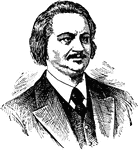
(1799-1850) French novelist and playwright most famous for Eugenie Grandet and La Comedie Humaine.
Honore de Balzac

(1799-1850) French novelist and playwright most famous for Eugenie Grandet and La Comedie Humaine.

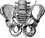
A bony structure located at the bottom of the spine. The human sacrum forms the back part of the pelvis, is roughly triangular in shape, consists of five united vertebrae, and from its solidity it is well adapted to serve as the keystone of the pelvic arch.

Pelvic Bone, Male

A bony structure located at the bottom of the spine. The human sacrum forms the back part of the pelvis,…

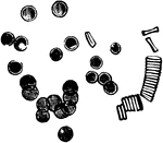
Human red blood corpuscles. Highly magnified.

Blood Corpuscles

Human red blood corpuscles. Highly magnified.

Chief veins and arteries of the body. Labels: a, place of the heart; the veins are in the back. On the right side of the picture the veins just under the skin are shown on the other side the deep vessels near the bones. b, vessels to the lungs.

Veins and Arteries of the Body

Chief veins and arteries of the body. Labels: a, place of the heart; the veins are in the back. On the…
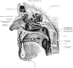
The nasal cavity with openings of accessory sinuses. The sagittal section has been made a little to the right of the nasal septum sphenoidal and frontal septum. Portions have been removed from the turbinated bones in order to exhibit the orifices of the accessory cavities. The tongue has been almost completely removed to show the tonsils.

Nasal Cavity with Openings of Accessory Sinuses

The nasal cavity with openings of accessory sinuses. The sagittal section has been made a little to…
The human skeleton.

The Human Skeleton

The human skeleton.

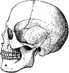
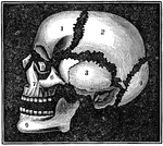
A diagram of the bones of the head. Label: 1, frontal lobe; 2, parietal bone; 3, temporal bone; 4, occipital bone; 5, nasal bone; 6, malar bone; 7, upper jaw; 8, os unguis; 9, lower jaw.

Bones of the Head

A diagram of the bones of the head. Label: 1, frontal lobe; 2, parietal bone; 3, temporal bone; 4, occipital…

The upper surface of the bones of the foot. Labels: 1, the surface of the astragulus or ankle bones, where it unites with the tibia. 2, The body of the astragulus. 3, Calcis or heel-bone. 4, The scaphoid. 5,6,7, The cuneiform. 8, The cuboid. 9,The metatarsal bones. 10,11, The phalanges of the great toe. 12, 13, 14, The phalanges of the other toes.

Bones of the Foot

The upper surface of the bones of the foot. Labels: 1, the surface of the astragulus or ankle bones,…

A diagram of the relative position of the bone, cartilage, and synovial membrane. Labels: 1,The extremities of two bones to form a joint. 2, The cartilage that covers the end of the bone. 3, The synovial membrane, which covers the cartilage of both bones and is then doubled back from one to the other, represented by the dotted lines.

Position of the Bone, Cartilage, and Synovial Membranes

A diagram of the relative position of the bone, cartilage, and synovial membrane. Labels: 1,The extremities…
A human skeleton from the back.

Back View of a Human Skeleton

A human skeleton from the back.

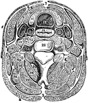
A transverse section of the neck. The separate muscles as they are arranged in layers, with their investing fascia. The figures in white space represent fascia (sheath); other figures are muscles. Labels: 12, trachea; 13, esophagus, 14, carotid artery and jugular vein; 28, one of the bones of the spinal column.

The Transverse Section of the Neck

A transverse section of the neck. The separate muscles as they are arranged in layers, with their investing…

The metacarpal and phalangeal bones of the fingers, with their tendons and ligaments. Labels: 1, metacarpal bone, 2. tendon of the superficial flexor. 3, Tendon of the deep flexor passing through a perforation (*) of the superficial flexor.

Metacarpal and Phalangeal Bones of the Fingers

The metacarpal and phalangeal bones of the fingers, with their tendons and ligaments. Labels: 1, metacarpal…

A diagram of the human muscular system.

A Diagram of the Human Muscular System

A diagram of the human muscular system.

The molar teeth of a human, horse and dog. The first image to the left in a molar tooth of a horse. Labels: 1, The enamel. 2, The ivory. 3, Canals for blood vessels. The center image is a molar tooth of a human. Label: 1, The enamel. 2, The ivory. 3, The cavity containing blood vessels. 4, 5, Artery and nerve. The image on the right is a molar tooth of a dog. Labels: 1, The enamel. 2, The ivory. 3, Cavity for blood-vessels.

Comparison of the Molar Teeth of a Human, Horse, and Dog

The molar teeth of a human, horse and dog. The first image to the left in a molar tooth of a horse.…
The human circulatory organs.

The Circulatory Organs

The human circulatory organs.

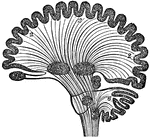
Diagram of human brain in vertical section, showing the situation of the different ganglia and the course of fibers. Labels: 1, Olfactory ganglion. 2, Hemisphere. 3, Corpus striatum. 4, Optic thalamus. 5, Tubercula quadrigemina. 6, Cerebellum. 7, Ganglion of tuber annulare. 8, Ganglion of medulla oblongata.

Diagram of Human Brain in Vertical Section

Diagram of human brain in vertical section, showing the situation of the different ganglia and the course…
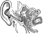
A view of all the parts of the ear, Labels: 1, The tube that leads to the internal ear. 2, The membrana tympani. 3, 4, 5, The bones of the ear. 7, The central part of the labyrinth, names the vestibule. 8, 9, 10, The semicircular canals. 11, 12, The channels of the cochlea. 13, The auditory nerve. 14, The channel from the middle ear to the throat (Eustachian tube). 15, A nerve. 16, A process. 17, The seventh pair of nerves (facial). 18, A process of the temporal bone.

Parts of the Ear

A view of all the parts of the ear, Labels: 1, The tube that leads to the internal ear. 2, The membrana…
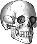
The human skull. Labels: 1, frontal lobe; 2, parietal lobe; 3, temporal lobe; 4, the sphenoid bone; 5, ethmoid bone; 6, superior maxillary (upper jaw) bone; 7, malar bone; 8, lachrymal bone; 9, nasal bone; 10, inferior maxillary (lower jaw) bone.

The Skull

The human skull. Labels: 1, frontal lobe; 2, parietal lobe; 3, temporal lobe; 4, the sphenoid bone;…

The human torso. Labels: A, the heart; B, the lungs drawn aside to show the internal organs; C, the diaphragm; D, the liver; E, the gall cyst; F, the stomach; G, the small intestines; H, the transverse colon.

Torso

The human torso. Labels: A, the heart; B, the lungs drawn aside to show the internal organs; C, the…
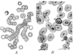
A magnified view of corpuscles of human blood compared to animal blood. Labels: A, corpuscles of human blood; B, corpuscles in the blood of an animal (a non-mammal).

Corpuscles of Human and Animal Blood

A magnified view of corpuscles of human blood compared to animal blood. Labels: A, corpuscles of human…

The human nervous system includes the brain, spinal cord, and the nerves. Labels: A, cerebrum; B, cerebellum.
Nervous System

The human nervous system includes the brain, spinal cord, and the nerves. Labels: A, cerebrum; B, cerebellum.

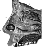
The olfactory system. Labels: a, b, c, d, interior of the nose, which is lined by a mucous membrane; n, the nose; e, the wing of the nose; q, the nose bones; o, the upper lip; g, section of the upper jawbone; h, the upper part of the mouth, or hard palate; m, frontal bone of the skull; k, the ganglion or bulb of the olfactory nerve in the skull, from which are seen the branches of the nerve passing in all direction.

Olfactory System

The olfactory system. Labels: a, b, c, d, interior of the nose, which is lined by a mucous membrane;…