Clipart tagged: ‘Abdominal’
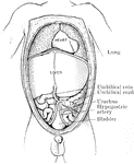
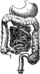
Abdomen of Fetus
The abdominal and thoracic viscera of a five months fetus. The large liver and large size if its left…

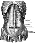
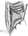
Muscles of the Abdomen
The muscles of the abdomen, showing the semilunar fold of Douglas. Viewed from in front.
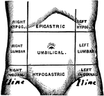
Regions of the Abdomen
Regions of the abdomen. The abdomen is divided into 9 regions by two horizontal planes, one at the level…
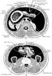
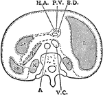
Transverse Section of Abdomen
Diagrammatic transverse section of abdomen, to show the peritoneum on transverse tracing. A, at level…
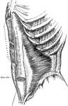
Transverse Section of the Abdomen
A transverse section of the abdomen in the lumbar region showing abdominal muscles.
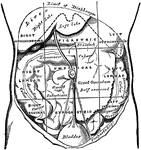
Abdominal Region
Showing the average position of the abdominal viscera with their surface markings. Labels: A, sterno-ensiform…
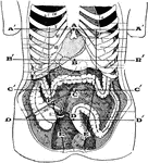
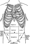
Regions of the Abdomen and their Contents
Regions of the abdomen and their contents (edge of costal cartilages in dotted outline). "For convenience…
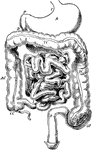
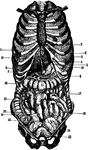
Alimentary Canal
Diagram of the abdominal part of the alimentary canal (digestive system). Labels: C, the cardiac, and…
Alimentary Canal
Diagram of the abdominal part of the alimentary canal. Labels: C, the cardiac, and P, the pyloric end…
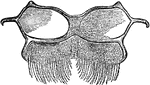
Bee Abdomen
"Abdominal Plate (worker of Apis), under side, third segment. W, wax-yielding surface, covering true…
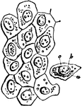
Cells from the Abdominal Lining
Flat cells from the surface of the lining membrane of the abdomen (peritoneum). Labels: a, cell-body;…
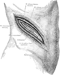
Surgical Incision to the Kidney
An incision in the right side above the kidney, showing a typical surgical approach to this organ, exposing…
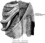
Transition of the Oblique Muscle into the Rectus
Transition of the tendon of the right internal oblique into the sheath of the rectus.

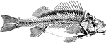
Perch Skeleton
"The spinal column consists of abdominal and caudal vertebre, the coalescence of the parapophyses into…
The Peritoneum
The peritoneum is a large serous membrane, which forms in the male a closed sac, the parietal layer…
Section Across the Forearm
Diagram showing the position of the thoracic and abdominal organs. labels: 1, lower border of right…
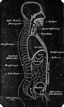
Thoracic and Abdominal Regions
Diagram of the thoracic and abdominal regions. Labels: A, aortic valve; P, pulmonary valve; M, mitral…
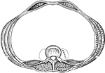
Horizontal Section Through Trunk
Diagram of horizontal section through upper part of 1st lumbar vertebra. The fine dots represent the…
Vertical Median Section of the Trunk
Diagram of vertical median section of abdomen. The fine dots represent the great sac of the peritoneum,…
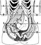
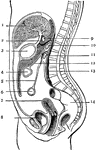
Position of the Viscera in the Condition of Visceroptosis
Showing the position of the viscera in the condition of visceroptosis (Glenard's disease). Labels: A,…