"The Main Nerve Trunks of the Right Forearm, showing the Accompanying Radial and Ulnar Arteries. (Anterior view.)" — Blaisedell, 1904

Nerve trunks

"The Main Nerve Trunks of the Right Forearm, showing the Accompanying Radial and Ulnar Arteries. (Anterior…

"A Great Nerve (Posterior Tibial) on the Back of the Leg, with its Accompanying Artery of the Same Name." — Blaisedell, 1904

Great Nerve

"A Great Nerve (Posterior Tibial) on the Back of the Leg, with its Accompanying Artery of the Same Name."…

"A Great Nerve (Crural) and its branches on the Front of the Thigh. The femoral artery with its cut end is shown." — Blaisedell, 1904

Great Nerve

"A Great Nerve (Crural) and its branches on the Front of the Thigh. The femoral artery with its cut…
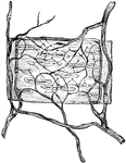
Ramifications of nerves and termination in muscular coat of a small artery of the frog.

Nerves in the Artery of a Frog

Ramifications of nerves and termination in muscular coat of a small artery of the frog.
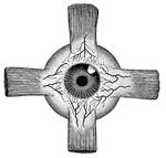
"Showing the attachment of the recti, or straight muscles to the eyeball, also the distribution of arteries upon the outer coat of the eye." — Blaisedell, 1904

Attachment of the recti

"Showing the attachment of the recti, or straight muscles to the eyeball, also the distribution of arteries…

"The renal organs, viewed from behind. R, right kidney; A, aorta; Ar, right renal artery; Vc, inferior vena cava; Vr, right renal vein; U, right ureter; Vu, bladder; Ua, commencement of urethra." —Martin, 1917

Renal Organs

"The renal organs, viewed from behind. R, right kidney; A, aorta; Ar, right renal artery; Vc, inferior…

This illustration shows a section of a human kidney (A, Cortical substance; B, Pyramids; C, Hilum; D, Pelvis; E, Ureter; F, Suprarenal capsule).
Kidneys are excretory organs whose function is to get rid of nitrogenous waste.

Section of Human Kidney

This illustration shows a section of a human kidney (A, Cortical substance; B, Pyramids; C, Hilum; D,…

"In the figure on the left are seen the artery and vein of a villus. In the right figure are represented the central lacteal of the villus, with the plexus of lymph vessels at the base. The mucous, submucuous, muscular, and serous coats are indicated at the base of the right-hand figure.  The epithelium of each villus is well shown." — Blaisedell, 1904

Transverse section of the small intestine

"In the figure on the left are seen the artery and vein of a villus. In the right figure are represented…
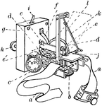
"An instrument which, when applied over an artery, traces on a piece of paper moved by clockwork a curve which indicates the changes of tension of the blood within... a, band by which the instrument is fastened on; b, spring which rests upon the artery; c, adjusting-screw (with graduated head) which regulates the pressure of the spring b according as the pulse is strong or weak; d, d, supports for paper upon which the tracing is made; e, e to engage the paper positively; g, small spring clockwork (incased) by which motion is imparted to the feed-roller e1; h, milled-headed winding key; i, stop-motion; j, tracer attached to the oscillating arm k, which is moved by the rod l that connects this arm with the spring b." —Whitney, 1889

Sphygmograph with All Parts Labeled

"An instrument which, when applied over an artery, traces on a piece of paper moved by clockwork a curve…
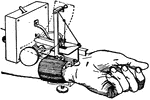
A device used for recording the movements of the arterial walls."—Finley, 1917

Dudgeon's sphygmograph

A device used for recording the movements of the arterial walls."—Finley, 1917

Internal view of left thigh-showing the arteries. Labels: 1, femoral; a, profunda femoris; b, superficialis femoris; c, saphenic; d, muscular branches; e, femora; nutrient vessels cut; (the femoral becomes the popliteal at about this point); f, femoro-popiteal; f', its muscular branches; f", its descending branch; g, recurrent tibial; h, united trunk of saphenic and femoro-popliteal; i, internal plantar.

Thigh of a Horse Showing Arteries

Internal view of left thigh-showing the arteries. Labels: 1, femoral; a, profunda femoris; b, superficialis…
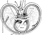
"Cross-section of thorax. A, bronchus, entering the lung; B, the aorta cut at its origin and again at the descending part of its arch; C, the pericardial space; D, the pleural cavity; E, the alimentary canal; PA, the pulmonary artery; X, the neural canal." —Martin, 1917

Thorax

"Cross-section of thorax. A, bronchus, entering the lung; B, the aorta cut at its origin and again at…

"Chief veins and arteries of the body. a, place of the heart; the veins are in black. On the right side of the pictures the veins just under the skin oare shown and on the other side the deep vessels near the bones.. b, vessels to the lungs." —Davison, 1910

Veins and arteries

"Chief veins and arteries of the body. a, place of the heart; the veins are in black. On the right side…

Chief veins and arteries of the body. Labels: a, place of the heart; the veins are in the back. On the right side of the picture the veins just under the skin are shown on the other side the deep vessels near the bones. b, vessels to the lungs.

Veins and Arteries of the Body

Chief veins and arteries of the body. Labels: a, place of the heart; the veins are in the back. On the…

"<em>A</em>, inferior vena cava, cut off just above the hepatic veins; <em>B</em>, superior vena cava, cut off just below the junction of the innominate veins; <em>C</em>, right and left innominate veins; <em>D</em>, left internal jugular vein. The veins, known as <em>azygos</em>, with some of their main branches, are shown. These veins connect the superior vena cava and the inferior vena cava." &mdash; Blaisedell, 1904

Vena cava

"A, inferior vena cava, cut off just above the hepatic veins; B, superior vena cava,…