Clipart tagged: ‘esophagus’
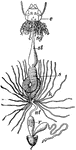
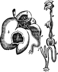
Ailmentary Canal
"ailmentary canal of a honey bee. at, honey stomach; s, true stomach; nt, intestine; o, esophagus; sg,…
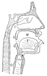
Passage to the Esophagus and Windpipe
The passage to the esophagus and windpipe. Labels: c, the tongue; d, the soft palate ending in g, the…
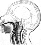
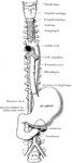
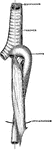
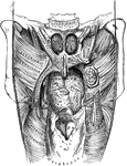
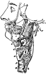
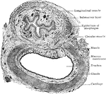
Position of the Esophagus in the Cervical Region
The position and relation of the esophagus in the cervical region and in the posterior mediastinum.…
Human Esophagus
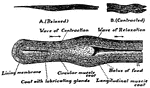
An Image of an esophagus showing how a mass of food, or bolus, passes through the esophagus into the…
Esophagus, Trachea, and Aorta in an Infant
The relationship of the esophagus, trachea and aorta in an infant.
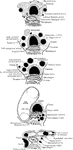
Development of the Intestinal Canal
Two diagrams to illustrate the development of the intestinal canal. The figure to the right shows the…
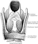
Larynx
The larynx viewed from its pharyngeal opening. The back wall of the pharynx has been divided and its…
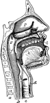
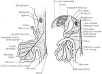
The Mouth, Nose, and Pharynx
The mouth, nose, and pharynx, with the commencement of gullet (esophagus) and larynx, as exposed by…
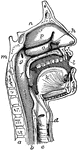
The Mouth, Nose, and Pharynx
The mouth, nose, and pharynx, with the larynx and commencement of gullet (esophagus), seen in section.…
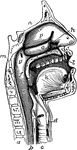
The Mouth, Nose, and Pharynx
The mouth, nose, and pharynx, with the commencement of the gullet (esophagus) and larynx, as exposed…
Nasal and throat passageways
"Diagram of a Sectional View of Nasal and Throat Passageways. C, nasal cavities; T,…
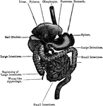
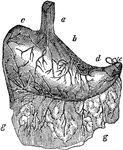
Ox Stomach
"Compound stomach of ox. a, esophagus; b, rumen, or paunch; c, reticulum, or second stomach; d, omasum,…
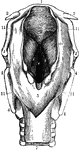
Muscles of the Soft Palate
Muscles of the soft palate, the pharynx being laid open from behind and mucous membrane removed.
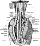
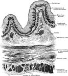
Pharynx and Esophagus
The lower part of the pharynx and the upper part of the esophagus have been slit up from behind, and…
Pleural Sac
Left pleural sac in a subject hardened by formalin injection, opened into by the removal of the costal…
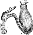
Shark Stomach
"Siphonal stomach and spiral valve of Basking-shark (Selache). a, esophagus; b, cardiac portion of stomach;…
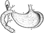
Stomach
The human stomach. Labels: a, the esophagus or gullet; b, the cardiac portion; c, the left extremity;…
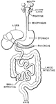
Vertical and Longitudinal Section of Stomach, Gall-Bladder, and Duodenum
Vertical and longitudinal section of stomach, gall-bladder, and duodenum. Labels: 1, esophagus; 2, cardiac…
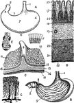
Views of the Stomach
Views of the stomach. Labels: A. stomach (human). B. Same, anterior wall removed. C. Portion of stomach,…
Swallowing Muscles
"The circular muscle of the mouth (1) and the buccinator or trumpeter's muscle (2) help the tongue to…
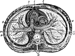
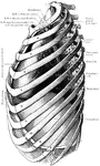
Thorax
"The transverse section of the thorax. 1, anterior mediastinum; 2, internal mammary vessels; 3, triangularis…
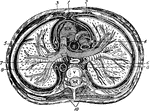
The Thorax
Diagram of a transverse section of the thorax. Labels: 1, anterior mediastinum; 2, internal mammary…
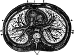
Transverse Section of the Thorax
Transverse section of the thorax. Labels: 1, anterior mediastinum; 2, internal mammary vessels; 3, triangularis…
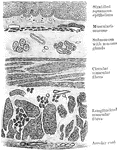
Transverse Section of Trachea and Esophagus
Transverse section of trachea and esophagus of child, seen from below.
Sections Through Trachea
Transverse section through the trachea and its immediate surroundings at the level of each of the upper…