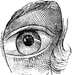
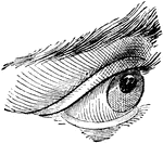
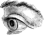
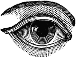
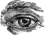
Clipart tagged: ‘eyeball’
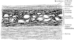
Vertical Section of the Chorioid and Sclera
Vertical section of the chorioid and inner part of the sclera.
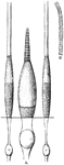
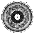
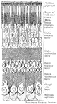
Cones and Rods of Retina
A. A cone and two rods from the human retina (modified from Max Schultze); B. Outer part of rod separated…
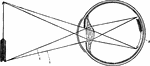
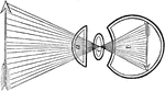
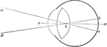
Convergence of Rays in the Aqueous Humor of the Eyeball
The convergence of light rays in the eyeball begins in the aqueous humor is perfected in the crystalline.…
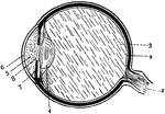
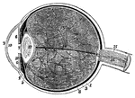
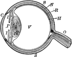
Eye
Section of the eye. 1: Optic nerve; 2: Retina; 3: Vitreous humor; 4: Crystalline lens; 5: Aqueous humor;…
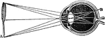
Eye Diagram
Diagram of the eye. 1: Lines of light from end of arrow; 2: Small, inverted image in the eye.
Human Eye
c, ciliary nerves going to be distributed in iris; d, smaller ciliary nerve; e, veins known as vasa…
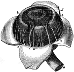
Human Eye
The iris and adjacent structures seen from behind. 1, the divided edge of the three coats, the choroid…
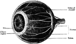
Human Eye
1, the sclerotic thicker behind than in front; 2, the cornea; 3, the choriod; 6, the iris; 7, the pupil;…
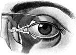
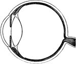
Eyeball
"The Relative Position of the Lachrymal Apparatus, the Eyeball, and the Eyelids. A, lachrymal canals,…
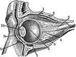
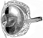
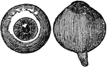
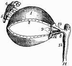
Eyeball
Section through the closed left eye. 1. Lifting muscle 2. Upper Straight Muscle 3. Optic Nerve 4. Fatty…
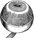
Eyeball
"The most essential parts of human vision are contained in the eyeball, a nearly spherical body, about…
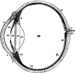
Left Eyeball in Horizontal Section
The left eyeball in horizontal section from before back. Labels: 1, sclerotic; 2, junction of sclerotic…
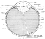
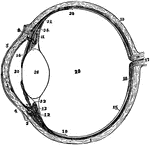
The Eyeball in Horizontal Section
The left eyeball in horizontal section from before back. Labels: 1, sclerotic; 2, junction of sclerotic…
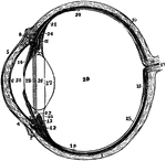
Section of Left Eyeball
The left eyeball in horizontal section from before back. Labels: 1, sclerotic; 2, junction of sclerotic…
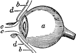
Side View of the Eyeball
Side view of the eyeball. Labels: a, the eyeball, and b,b, are the upper and lower sides. Now in order…
Formation of an Image on the Eyeball
In passing through the crystalline, the rays cross each other, so that those rays which pass from the…
Section Through Lens
Section through the equator of the lens. Showing gradual transition of the epithelium into lens fibers.
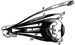
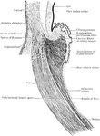
Eye Muscles
The muscles of the eyeball, the view being taken from the outer side of the right orbit.
Eye Muscles
1, cartilage of the upper eyelid; 2, its lower border, showing the openings of the Meibomian glands;…
Attachment of the recti
"Showing the attachment of the recti, or straight muscles to the eyeball, also the distribution of arteries…
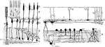
Section of Retina
Perpendicular section of mammalian retina. Labels: A, layer of rods and cones; B, outer nuclear layer;…