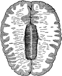
Cross-section of the brain. Here the upper half of the brain is cut off, and you see the upper cut surface of the lower half. The outer shaded part is the gray matter, and the inner lighter area is the white matter. In the center is the corpus callosum, a type of white matter that connects the two hemispheres together.

Cross-Section of the Brain

Cross-section of the brain. Here the upper half of the brain is cut off, and you see the upper cut surface…

Longitudinal and vertical diagrammatic section of a vertebrate brain. Mb, midbrain: what lies in front of this is the fore-, and what lies behind, the hindbrain; Lt, lamina terminalis; Olf, olfactory lobes; Hmp, hemispheres; Th. E, thalamencephalon; Pn, pineal gland; Py, pituitary body; F. M., foramen of Munro; cs, corpus striatum; Th, optic thalamus; CC, crura cerebri; the mass lying above the canal represents the corpora quadrigemina; Cb, cerebellum; I-IX, the nine pairs of cranial nerves; 1, olfactory ventricle; 2, lateral ventricle; 3, third ventricle; 4, fourth ventricle; +, iter a tertio ad quartum ventriculum. Lamina terminalis is represented by the strong black line joining Pn and Py.

Vertical Section of a Vertebrate Brain

Longitudinal and vertical diagrammatic section of a vertebrate brain. Mb, midbrain: what lies in front…

The five layers of the cortical gray matter of the cerebrum. 1, Superficial layer with abundance of neuroglia and few small multipolar ganglion cells. 2, A thin layer of a large number of closely packed small ganglion cells of pyramidal shape. 3, The most important layer, and the thickest of all: it contains many large pyramidal ganglion cells, each with a process running off from the apex vertically towards the free surface, and lateral processes at the base which are always branched. 4, Numerous ganglion cells come are large and others small, forming the granular formation of Meynert. 5, Spindle-shaped and branched ganglion cells of moderate size arranged chiefly parallel to the free surface.

Cortical Gray Matter of the Cerebrum

The five layers of the cortical gray matter of the cerebrum. 1, Superficial layer with abundance of…
Section through the gray matter of the human cerebellum.

Gray Matter of Cerebellum

Section through the gray matter of the human cerebellum.

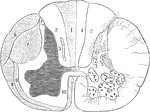
Section of a spinal cord, one half of which shows the tracts of the white matter, and the other half (right) shows the position of the nerve cells in the gray matter. 7, 10, 9, and 3 are tracts of descending degeneration, 1, 4, 6, and 8, of the ascending degeneration.

Section of the Spinal Cord

Section of a spinal cord, one half of which shows the tracts of the white matter, and the other half…
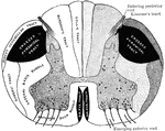
Diagrammatic representation of a transverse section through the spinal cord. The nerve tracts in the white matter and the clusters of the nerve cells in the gray matter are shown.

Transverse Section Through Spinal Cord

Diagrammatic representation of a transverse section through the spinal cord. The nerve tracts in the…