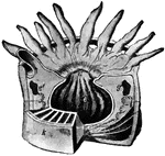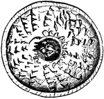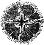
"The under surface, showing the mouth in the center, surrounded by the tentacula, and overial chambers exterior to the origins of these." — Chambers' Encyclopedia, 1875

Acalephae

"The under surface, showing the mouth in the center, surrounded by the tentacula, and overial chambers…
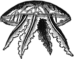
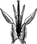
"Side-view, showing the tentacula hanging down in their natural position." — Chambers' Encyclopedia, 1875

Acalephae

"Side-view, showing the tentacula hanging down in their natural position." — Chambers' Encyclopedia,…

"The head is represented as having been thrown back, and the tongue drawn forward. <em>A</em>, <em>B</em> and <em>C</em>, cainine; <em>D, E</em> bicuspids; <em>F, H, K,</em> molars; <em>M</em> anterior pillar of the fauces; <em>N</em>, tonsil; <em>L</em> uvula; <em>O</em>, upper part of the pharynx; <em>P</em>, tongue drawn forward; <em>R</em> linear ridge, or raphe</em>." &mdash; Blaisedell, 1904

Back view of the adult mouth

"The head is represented as having been thrown back, and the tongue drawn forward. A, B

"Sea anemone dissected; c, tentacles; d, mouth; e, stomach; white lines above k, the mesenteries." &mdash;Davison, 1906

Anemone

"Sea anemone dissected; c, tentacles; d, mouth; e, stomach; white lines above k, the mesenteries." —Davison,…
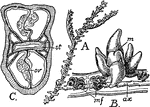
"A, portion of a colony of Antipathes dichotoma. B, single zooid and axis of the same magnified. m, mouth; mf, mesenterial filament; ax, axis. C, transverse section through the oral cone of Antipathella minor; st, stomodaeum; ov, ovary." &mdash; The Encyclopedia Britannica, 1910

Antipathes Dichotoma

"A, portion of a colony of Antipathes dichotoma. B, single zooid and axis of the same magnified. m,…

The bill or nib of a bird, turtle, etc.

Beak

The bill or nib of a bird, turtle, etc.

A decorative tailpiece with a horned beast with its mouth open in the center.

Beast Tailpiece

A decorative tailpiece with a horned beast with its mouth open in the center.

A worker bee's mouth. <em>a, e, f</em> tongue and ducts; <em>b</em> labial palps; <em>c</em> paraglossae; <em>d</em> first maxillae.

Bee Mouth

A worker bee's mouth. a, e, f tongue and ducts; b labial palps; c paraglossae;…

"The structure of the mouth in insects exhibits very remarkable modifications, and these are of the utmost importance in the classification of these creatures. In some insects the mouth is formed exclusively for biting; in others, as exclusively for suction; while in others again it is fitted for the performance of both these actions; and the form of its constituent parts of course undergoes corresponding changes; but the same organs really exist in all, modified in apearance, indeed, so as sometimes to be scarecely recognizable." &mdash; Goodrich, 1859

Mouth and Tongue of a Bee

"The structure of the mouth in insects exhibits very remarkable modifications, and these are of the…
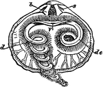
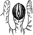
"a, tentacula; b, mouth; c, termination of intestine." &mdash; Chambers' Encyclopedia, 1875

Beroe

"a, tentacula; b, mouth; c, termination of intestine." — Chambers' Encyclopedia, 1875

The beak of a fowl.

Bill

The beak of a fowl.

A diagram of the digestive system.

Digestive System

A diagram of the digestive system.

"Rhynchonella psittacea. Interior of doral valve. s, sockets; b, dental plates; V, mouth; de, labial appendage in itsnatural position; d, appendage extended or unrolled." &mdash; Encyclopedia Britanica, 1893

Dorsal Valve

"Rhynchonella psittacea. Interior of doral valve. s, sockets; b, dental plates; V, mouth; de, labial…
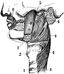
A side view of face. 1 and 2: Trachea. 3: Esophagus. 4, 5, and 6: Muscles. 7: Submaxillary. 8: Parotid gland. 9: Duct from the Parotid gland.

Face

A side view of face. 1 and 2: Trachea. 3: Esophagus. 4, 5, and 6: Muscles. 7: Submaxillary. 8: Parotid…
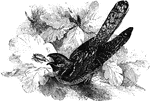
A small bird with a large mouth, which preys primarily on small insects.

European Goat-Sucker

A small bird with a large mouth, which preys primarily on small insects.

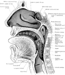
Sagittal section through mouth, tongue, larynx, pharynx, and nasal cavity. The section was slightly oblique, and the posterior edge of the nasal septum has been preserved. The specimen is viewed slightly from below, hence the apparently low position of the inferior turbinated bone.

Section of the Head and Neck

Sagittal section through mouth, tongue, larynx, pharynx, and nasal cavity. The section was slightly…

This illustration shows an enlarged view of the mouth of a lamprey.

Mouth of Lamprey

This illustration shows an enlarged view of the mouth of a lamprey.

"Diagram of a lateral view of a longitudinal section of Limulus. Suc, Suctorial pharynx. al, Alimentary canal. Ph, Pharynx. M, Mouth. Est, Entosternum. VS, Ventral venous sinus. chi, Chilaria. go, Genital operculum. br1 to br5, Branchial appendages. met, Unsegmented metasoma. Entap4, Fourth dorsal entapophysis of left side. tsm, Tergo-sternal muscles, six pairs as in Scorpio. VPM1 to VPM8, The eight pairs of veno-pericardiac muscles." &mdash; The Encyclopedia Britannica, 1910

Limulus

"Diagram of a lateral view of a longitudinal section of Limulus. Suc, Suctorial pharynx. al, Alimentary…

"View of the ventral surface of the mid-line of the prosomatic region of Limulus polyphemus. The coxae of the five pairs of limbs following the chelicerae were arranged in a series on each side between the mouth, M, and the metasternites, mets. sf, The sub-frontal median sclerite. Ch, the chelicerae. cam, The camerostome or upper lip. M, the mouth. pmst, The promesosternal sclerite or chitinous plate, unpaired. mets, the right and left metasternites (corresponding to the similarly placed pentagonal sternite of Scorpio." &mdash; The Encyclopedia Britannica, 1910

Limulus Polyphemus

"View of the ventral surface of the mid-line of the prosomatic region of Limulus polyphemus. The coxae…

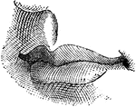
Lips

Lips

Lips

Lips

Lips

Lips

Lips

Lips

Lips

Lips

Lips

Lips

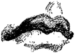
The lips, nose, and chin of a person's face.

Lips

The lips, nose, and chin of a person's face.

Head of female mosquito, showing the piercing needle-like mouth parts.

Mosquito

Head of female mosquito, showing the piercing needle-like mouth parts.

"The mouth, nose and pharynx, with the commencement of the gullet and larynx, as exposed by a section, a little to the left of the median plane of the head. a, vertebral column; b, gullet; c, wind-pipe; d, larynx; e, epiglottis; f, soft palate; g, opning of eustachian tube; k, tongue; l, hard palate; m, the sphenoid bone on the base of the skull; n, the fore part of the cranial cavity; o, p, q, the turbinate bones of the outer side of the left nostril-chamber." &mdash;Martin, 1917

Mouth

"The mouth, nose and pharynx, with the commencement of the gullet and larynx, as exposed by a section,…
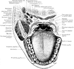
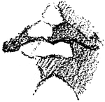
A mouth

Mouth

A mouth

The soft palate and tonsillar regions.

Mouth

The soft palate and tonsillar regions.

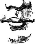
A sketch of the human mouth and chin.

Mouth and Chin

A sketch of the human mouth and chin.

A sketch of the human mouth and chin.

Mouth and Chin

A sketch of the human mouth and chin.

A sketch of the human mouth and chin.

Mouth and Chin

A sketch of the human mouth and chin.

A sketch of the human mouth and chin.

Mouth and Chin

A sketch of the human mouth and chin.

A sketch of the human mouth and chin.

Mouth and Chin

A sketch of the human mouth and chin.

A sketch of the human mouth and chin.

Mouth and Chin

A sketch of the human mouth and chin.

A sketch of the human mouth and chin.

Mouth and Chin

A sketch of the human mouth and chin.

Horizontal section through mouth and pharynx at the level of the tonsils.

Horizontal Section Through Mouth and Pharynx

Horizontal section through mouth and pharynx at the level of the tonsils.
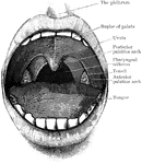
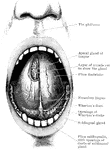
Open mouth with tongue raised, and the sublingual and apical glands exposed.

Mouth and Tongue

Open mouth with tongue raised, and the sublingual and apical glands exposed.

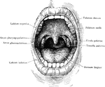
Open mouth showing palate and tonsils. It also shows the two palatine arches, and the pharyngeal isthmus through which the nasopharynx above communicates with the oral portion of the pharynx below.

Mouth Showing Palate and Tonsils

Open mouth showing palate and tonsils. It also shows the two palatine arches, and the pharyngeal isthmus…

Section across anterior part of floor of mouth, showing relations of sublingual glands to mucous membrane and muscles.

Anterior Part of Floor of Mouth

Section across anterior part of floor of mouth, showing relations of sublingual glands to mucous membrane…
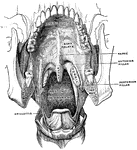
Antero-inferior surface of the soft palate. The tongue has been removed, so that the pharyngeal isthmus is distinctly seen.

Mouth, Cavity of the

Antero-inferior surface of the soft palate. The tongue has been removed, so that the pharyngeal isthmus…

The mouth, nose, and pharynx, with the commencement of gullet (esophagus) and larynx, as exposed by a section a little to the left of the median plane of the head. Labels: a, vertebrae; b, gullet; c, trachea; d, larynx; e, epiglottis; f, soft palate; g, opening of Eustachian tube; k, tongue; l, hard palate; o, p, q, inferior turbinate bones of left nasal chamber.

The Mouth, Nose, and Pharynx

The mouth, nose, and pharynx, with the commencement of gullet (esophagus) and larynx, as exposed by…

The mouth, nose, and pharynx, with the larynx and commencement of gullet (esophagus), seen in section. Labels: a, vertebral column; b, gullet (esophagus); c, trachea; d, larynx; e, epiglottis; f, soft palate, between f and e is the opening at back of cavity or faces; g, opening of Eustachian tube; h, nasal cavity; k, tongue; l, hard palate; m, sphenoid bone at base of skull; n, roof of nasal cavity; o, p, q, placed in nasal cavity.

The Mouth, Nose, and Pharynx

The mouth, nose, and pharynx, with the larynx and commencement of gullet (esophagus), seen in section.…

The mouth, nose, and pharynx, with the commencement of the gullet (esophagus) and larynx, as exposed by a section, a little to the left of the median plane of the head. Labels: a, vertebral column; b, gullet; c, windpipe; d, larynx; e, epiglottis; f, soft palate; g, opening of Esutachian tube; k, tongue; l, hard palate; m; the sphenoid bone on the base of the skull; n, the fore part of the cranial cavity; o,p,q, the turbinate bones of the outer side of the left nostril chamber.

The Mouth, Nose, and Pharynx

The mouth, nose, and pharynx, with the commencement of the gullet (esophagus) and larynx, as exposed…

Coronal section through the closed mouth. The slit liked character of the vestibule, the manner which the tongue fills up the mouth cavity, the close apposition of the teeth, the relations of the roots of the upper molars to the antrum of Highmore, the plica sublingualis over the sublingual gland, and the position of the ranine artery should be noted.

Section Through Mouth

Coronal section through the closed mouth. The slit liked character of the vestibule, the manner which…
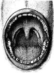
Interior of the mouth. Labels: 1, soft palate; 2, its median ridge; 3, uvula; 4, anterior, 5, posterior pillar of fauces; 6, tonsil; 7, posterior wall of pharynx; 8, tongue.

The Mouth

Interior of the mouth. Labels: 1, soft palate; 2, its median ridge; 3, uvula; 4, anterior, 5, posterior…

"Diagram showing the muscular system. M, ventral, N, dorsal valve; l, loop; V, mouth; Z, extremity of intestine; a, adductor; c, divaricators; c', accessory divaricators; b, ventral adjustors; c, peduncular muscles; b''', dorsal adjustors; P, peduncle." &mdash; Encyclopedia Britanica, 1893

Muscular System

"Diagram showing the muscular system. M, ventral, N, dorsal valve; l, loop; V, mouth; Z, extremity of…

"Diagram of a Sectional View of Nasal and Throat Passageways. <em>C</em>, nasal cavities; <em>T</em>, tongue; <em>L</em>, lower jaw; <em>M</em>, Mouth; <em>U</em>, uvula; <em>E</em>, epiglottis; <em>G</em>, larynx; <em>O</em>, gullet, or esophagus." &mdash; Blaisedell, 1904

Nasal and throat passageways

"Diagram of a Sectional View of Nasal and Throat Passageways. C, nasal cavities; T,…
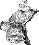
The oral cavity seen from in front.

Oral Cavity

The oral cavity seen from in front.

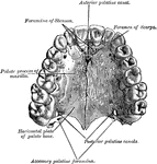
The palate and alveolar arch.

Palate

The palate and alveolar arch.

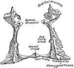
Palate bone. Palate bones form the back part of the roof of the mouth; part of the floor and outer wall of the nasal fossae, and a very small portion of the floor of the orbit.

Human Palate Bone

Palate bone. Palate bones form the back part of the roof of the mouth; part of the floor and outer wall…

The two palate bones in their natural position.

Palate Bones

The two palate bones in their natural position.

An illustration of the mouth of the Miami River. The Miami River is a river in Florida that drains out of the Everglades and runs through downtown Miami, Florida. The 5.5 mile (8.9 km) long river flows from the terminus of the Miami Canal at Miami International Airport to Biscayne Bay. It was originally a natural river inhabited at its mouth by the Tequesta Indians, but it was dredged and is now polluted because of its proximity to the Miami-Dade County. The mouth of the river is now home to the Port of Miami and many other businesses whose pressure to maintain it has helped to improve the river's condition.

Miami River

An illustration of the mouth of the Miami River. The Miami River is a river in Florida that drains out…
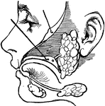
"There are three pairs of salivary glands. One pair lies under the tongue; one pair is found under the corners of the lower jaw; and the other pair is found in front of and below the ears." &mdash; Ritchie, 1918

Salivary glands

"There are three pairs of salivary glands. One pair lies under the tongue; one pair is found under the…

"Ventral view of a scorpion. Palamnaeus indus, de Geer, to show the arrangement of the coxae of the limbs, the sternal elements, genital plate and pectens. M, Mouth behind the oval median camerostome. I, The chelicerae. II, The chelae. III to VI, the four pairs of walking legs. VIIgo, The genital somite or first somite of the mesosoma with the genital operculum (a fused pair of limbs). VIIIp, the pectiniferous somite. IXstg to XIstg, the four pulmonary somites. met, the pentagonal metasternite of the prosoma behind all the coxae. x, the sternum of the pectiniferous somite. y, the broad first somite of the metasoma." &mdash; The Encyclopedia Britannica, 1910

Scorpion

"Ventral view of a scorpion. Palamnaeus indus, de Geer, to show the arrangement of the coxae of the…

"Diagram of a lateral view of a longitudinal section of a scorpion. d, Chelicera. ch, Chela. cam, Camerostome. m, Mouth. ent, Entosternum. p, Pecten. stig1, First pulmonary aperture. stig4, Fourth pulmonary aperture. dam, Muscle from carapace to a praeoral entosclerite. ad, Muscle from carapace to entosternum. md, Muscle from tergite of genital somite to entosternum. dv1 to dv6, Dorso-ventral muscles. pv1 to pv7, The seven veno-pericardiac muscles of the right side." &mdash; The Encyclopedia Britannica, 1910

Scorpion

"Diagram of a lateral view of a longitudinal section of a scorpion. d, Chelicera. ch, Chela. cam, Camerostome.…
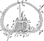
"Diagram of an Echinus (stripped of its spines). a, mouth; a', gullet; b, teeth; c, lips; d, alveoli; e, falces; f, f, auriculariae; g, retractor, and h, protractor, muscles of Aristotle's lantern; i, madreporic canal; k, circular ambulacral vessel; l, Polian pedicels; r, r, spines; s, tubercle; s', tubercle to which a spine is articulated; t, t, pedicellariae; u, anus; v, madreporic tubercle; x, ocular spot." -Whitney, 1911

Sea Urchin Section

"Diagram of an Echinus (stripped of its spines). a, mouth; a', gullet; b, teeth; c, lips; d, alveoli;…
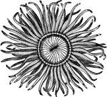
"Their tentacles, which are disposed in regular circles, and tinged with a variety of bright lively colors, very nearly represent the beautiful petals of some f the most elegantly fringed and radiated flowers, such as the carnation, marygold, and anemone. They are of various sizes, from that of the smallest thimble to the largest apple; and have considerable power of locomotion, being able not only to move along upon the base, but also in a reversed position upon their entacles." &mdash; Goodrich, 1859

Mouth of the sea-anemone

"Their tentacles, which are disposed in regular circles, and tinged with a variety of bright lively…

"Anatomy of the Snail: a, the mouth; bb, foot; c, anus; dd, lung; e, stomach, covered above by the salivary glands; ff, intestine; g, liver; h, heart; i, aorta; j, gastric artery; l, hepatic artery; k, artery of the foot; mm, abdominal cavity, supplying the place of a cenous sinus; nn, irregular canal in communication with the abdominal cavaty, and carrying the blood to the lung; oo, vessel carrying the blood from the lung to the heart." &mdash; Chambers, 1881

Snail Anatomy

"Anatomy of the Snail: a, the mouth; bb, foot; c, anus; dd, lung; e, stomach, covered above by the salivary…

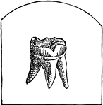
A small bone attached to the jaw for chewing.

Tooth

A small bone attached to the jaw for chewing.

Diagrammatic longitudinal section of the trunk and head. Labels: 1,1, the dorsal cavity; a, the spinal portion; b, the cranial enlargement; c, c, the bodies of the vertebrae forming the partition between the dorsal and ventral cavities; 2, 2, the ventral cavity, subdivided into thoracic cavity (d), abdominal cavity (e), and pelvic cavity (f); g, the nasal cavity; h, the mouth, or buccal cavity. The alimentary canal (al) is represented running through the whole length of the ventral cavity.

Trunk and Head of Human Body

Diagrammatic longitudinal section of the trunk and head. Labels: 1,1, the dorsal cavity; a, the spinal…