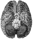
<em>A</em>, anterior lobe of the cerebellum; <em>B</em>, olfactory nerve; <em>C</em>, portion of the posterior lobe; <em>D</em>, optic chiasm; <em>E</em>, optic tract; <em>H, M,</em> hemispheres of the cerebellum; <em>K</em>, portion of the occipital lobe; <em>N</em>, medulla oblongata; <em>R</em>, pons Varolii."

Base of the brain

A, anterior lobe of the cerebellum; B, olfactory nerve; C, portion of the…

A simple nerve cell, or neuron. N is the nucleus of the cell, NC is the cytoplasm, D are dendrites which are fine protoplasmic branches that create synapses that connect to another nerve cell. The neuron is connected to muscle (MU) by a nerve fiber (NF). The fiber is covered by a sheath (SH) of fat to protect it. NE is the ending of the fiber that the muscle is connected to. LB is a lateral branch of the nerve fiber.

Nerve Cell

A simple nerve cell, or neuron. N is the nucleus of the cell, NC is the cytoplasm, D are dendrites which…
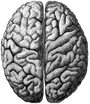
"The Upper Surface of the Cerebrum. Showing its division into two hemispheres, and also of the convolutions." &mdash; Blaisedell, 1904

Cerebrum

"The Upper Surface of the Cerebrum. Showing its division into two hemispheres, and also of the convolutions."…

Diagram of a neuron.

The Nervous System

Diagram of a neuron.

Diagram of nervous system. Labels: a, a, cortex of cerebral hemispheres; b, b, cell body and dendrites of upper motor neuron, situated in cerebral cortex; b', axon of upper motor neuron, branching at its termination near the dendrites of lower motor neuron, situated in the ventral horn of gray matter in the spinal cord; B', axon of lower motor neuron passing to its termination in a voluntary muscle fiber B"; C, cell body and dendrites of supper sensory neuron, situated in the medulla oblongata; C'C', axons of upper sensory neurons, terminating in cortex; c, cell body of lower sensory neuron situated in the dorsal root ganglion; c''', dendrite of lower motor neuron, conducting impulses from the periphery to the central nervous system; c", long axon of lower sensory neuron, conducting impulses toward the brain; c', short axon of lower sensory neuron, conducting impulses direct to ventral horn. (For the sake of simplicity the connection with the cerebellum are omitted.)

Nervous System Diagram

Diagram of nervous system. Labels: a, a, cortex of cerebral hemispheres; b, b, cell body and dendrites…

"Showing a motor cell with its long, unbranched process (with two little lateral offshoots), with motor endings in striated, voluntary muscular tissue." &mdash; Blaisedell, 1904

Neuron

"Showing a motor cell with its long, unbranched process (with two little lateral offshoots), with motor…

"A nerve cell from the brain." &mdash; Ritchie, 1918

Neuron

"A nerve cell from the brain." — Ritchie, 1918

A neuron or nerve cell.

Neuron

A neuron or nerve cell.

Diagram of typical neuron.

Neuron

Diagram of typical neuron.

Diagram of nerve cell of type II, in which axone is not prolonged as nerve fiber.

Neuron

Diagram of nerve cell of type II, in which axone is not prolonged as nerve fiber.

Diagram of a neuron. Labels: A, axon arising from the cell-body and branching at its termination; D, dendrites; C and N, cell-body composed of C, cytoplasm, and N, nucleus.

Diagram of a Neuron

Diagram of a neuron. Labels: A, axon arising from the cell-body and branching at its termination; D,…
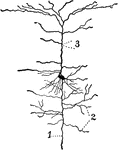
A neuron from the cerebral cortex. The axis-cylinder process, dendrites and collaterals are marked 1, 2, and 3, respectively.

Neuron from the Cerebral Cortex

A neuron from the cerebral cortex. The axis-cylinder process, dendrites and collaterals are marked 1,…
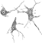
Nerve cells of human spinal cord stained to show Nissl bodies. Labels: D, dendrites; A, axons; C, implantation cone; N, nucleus; M, nucleolus.

Neuron of Spinal Cord

Nerve cells of human spinal cord stained to show Nissl bodies. Labels: D, dendrites; A, axons; C, implantation…

Semidiagrammatic representation of structure of a neuron; a, axon.

Structure of a Neuron

Semidiagrammatic representation of structure of a neuron; a, axon.

Showing some varieties of cell bodies of neurons. A, Unipolar (amacrine) cell from retina; B, Bipolar cell from vestibular ganglion; C, Multipolar cell, with long axon, from spinal cord; D, Golgi cell with short axon breaking up into numerous terminal twigs; E, pyramidal cell from cerebral cortex; a, Axon; clt., collaterals; t, telodendria.

Variety of the Cell Bodies of Neurons

Showing some varieties of cell bodies of neurons. A, Unipolar (amacrine) cell from retina; B, Bipolar…

Multipolar nerve cells of various forms. Labels: A, from spinal cord; B, from cerebral cortex; C, from cerebellar cortex (Purkinje cell); a, axon; c, implantation cone.

Various Forms of Neurons

Multipolar nerve cells of various forms. Labels: A, from spinal cord; B, from cerebral cortex; C, from…
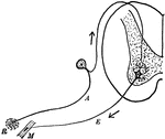
"Diagram of the simple reflex arc. R, receptor; A, afferent (sensory) neuron; E, efferent (motor) meuron; M, muscle." &mdash;Martin, 1917

Reflex Arc

"Diagram of the simple reflex arc. R, receptor; A, afferent (sensory) neuron; E, efferent (motor) meuron;…
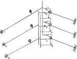
"Diagram to illustrate how a single sensory neuron may communicate with several motor neurons, and a single motor neuron with several sensory neurons." &mdash;Martin, 1917

Sensory Neuron

"Diagram to illustrate how a single sensory neuron may communicate with several motor neurons, and a…