"Profile and vertex views of cerebrum. Dr, the frontal lobe; Par, parietal; Oc, occipital; Ts, temporo-sphenoidal lobe; SS, Sylvian fissure; RR, fissure of Rolando; PO, parieto-occipital fissure; IP, intra-pariental fissure; PP, Parallel fissure; SF and IF, supero- and infero-frontal fissures; 1, inferior, 2 middle, and 3, superior frontal convolutions; 4, ascending frontal convolution; 5, ascending parietal, 5', postero-parietal, and 6, angular convolutions; A, supra-marginal, or convolution of the parietal eminence; 7, superior, 8, middle, and 9, inferior temporo-sphenoidal convolutions; 10, superior, 11, middle, and 12, inferior occipital convolutions." — Encyclopedia Britanica, 1893

Brain

"Profile and vertex views of cerebrum. Dr, the frontal lobe; Par, parietal; Oc, occipital; Ts, temporo-sphenoidal…
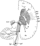
Diagram to show the connecting of the Frontal Occipital Lobes with the Cerebellum. The dotted lines passing in the crusta, outside the motor fibers, indicate the connection between the temporo-occipital lobe and the cerebellum. F.C., the fronto-cerebellar fibers, which pass internally to the motor tract in the crusta; I.F., fibers from the caudate nucleus to the pons. FR., frontal lobe; Oc, occipital lobe; AF. ascending frontal; AP., ascending frontal convolution; FR., fissure of Rolando; IPF., interparietal fissure, a section of crus is lettered on the left side. SN., substantia nigra; PY. pyramidal motor fiber, which on the right is shown as continuous lines converging to pass through the posterior limb of IC. internal capsule ( the knee or elbow of which is shown thus) upwards into the hemisphere and downwards through the pons to cross at the medulla in the anterior pyramids.

Brain Showing Connection of Frontal Occipital Lobe with Cerebellum

Diagram to show the connecting of the Frontal Occipital Lobes with the Cerebellum. The dotted lines…
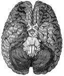
<em>A</em>, anterior lobe of the cerebellum; <em>B</em>, olfactory nerve; <em>C</em>, portion of the posterior lobe; <em>D</em>, optic chiasm; <em>E</em>, optic tract; <em>H, M,</em> hemispheres of the cerebellum; <em>K</em>, portion of the occipital lobe; <em>N</em>, medulla oblongata; <em>R</em>, pons Varolii."

Base of the brain

A, anterior lobe of the cerebellum; B, olfactory nerve; C, portion of the…

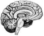
"<em>A</em>,frontal love of the cerebrum; <em>B</em>, parietal lobe; <em>C</em>, parieto-occipital lobe; <em>D</em>, occcipital lobe; <em>E</em>, cerebellum; <em>F</em>; arbor vitae; <em>H</em>, pons Varolii; <em>K</em>, medulla oblongata." — Blaisedell, 1904

Left Half of the Brain

"A,frontal love of the cerebrum; B, parietal lobe; C, parieto-occipital lobe;…

"Brain of chimpanzee. Ol, olfactory lobe; A, B, C, frontal, occipital, and temporal lobes; C1, a portion of temporal lobe; Sy, Sylvian fissure; In, insula or island of Reil; SOr, supra-orbital gyrus; SF, MF, IF, superior, middle, and inferior frontal gyri; R, fissure of Rolando; PPl, posteroparietal lobule; OPf, occipitotemporal sulcus; An, angular gyrus; 2, 3, 4, annectent gyri; AT, MT, PT, the anterior, middle, and posterior temporal gyri; SOc, MOc, IOc, the superior, middle, and inferior occipital gyri." -Whitney, 1911

Chimpanzee Brain

"Brain of chimpanzee. Ol, olfactory lobe; A, B, C, frontal, occipital, and temporal lobes; C1, a portion…
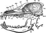
"Longitudinal and Vertical section of the skull of a dog, with mandible and hyoid arch. an, anterior narial aperture; MT, maxillo-turbinal bone; ET, the ethmo-turbinal; Fr, frontal; Pa, pariental; IP, interparietal; SO, supra-occipital; ExO, sphenoid; OS, orbito-sphenoid; PS, presphenoid; Pl, palatine; Vo, vomer; Mx, maxilla; PMx, premaxilla; sh, stylo-hyal; eh, epi-hyal; ch, cerato-hyal; bh, basihyal; th, thyro-hyal; s, symphysis of mandible; cp, coronoid process; cd, condyle; a, angle; id, inferior dental canal. The mandible is displaced downwards, to show its entire form; the * indicated the part of the cranium to which the condyle is articulated." &mdash;The Encyclopedia Britannica, 1903

Dog Skull

"Longitudinal and Vertical section of the skull of a dog, with mandible and hyoid arch. an, anterior…
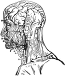
"1, primitive carotid artery dividing itself into carotid external and carotid internal; 3, occipital branch to the back part of the skull; 4, upper hyoides artery; 5, lower pharyngeal artery; 6, masseter artery; 7, submental artery; 8, lower coronary artery; 9, upper coronary artery; 10, deep branch; 11, back cervical artery; 12, continuation and fold of the occipital; 13, descending branch for muscles of the neck; 14, posterior auricular; 15, temporal artery; 16, pariental branches; 17, frontal branches." &mdash; Richardson, 1906

Facial Arteries

"1, primitive carotid artery dividing itself into carotid external and carotid internal; 3, occipital…
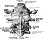
Occipital bone and first three cervical vertebrae with ligaments, from in front.

Occipital Bone and Cervical Vertebrae

Occipital bone and first three cervical vertebrae with ligaments, from in front.
Occipital bone at birth.

Occipital Bone at Birth

Occipital bone at birth.

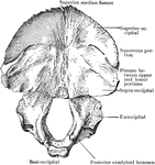
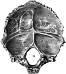
Occipital bone of the human skull, inner surface. It is situated at the back and base of the skull. Labels: 9,9, and 10,10, depression for reception of lobes of brain; 11, foramen magnum.

Occipital Bone of the Human Skull

Occipital bone of the human skull, inner surface. It is situated at the back and base of the skull.…

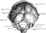
Occipital bone, internal surface, from before.

Internal Surface of the Occipital Bone

Occipital bone, internal surface, from before.

"Brain of pig. Ol, olfactory lobe; A, B, C, frontal, occipital, and temporal lobes; C1, a portion of temporal lobe; Sy, Sylvian fissure; In, insula or island of Reil." -Whitney, 1911

Pig Brain

"Brain of pig. Ol, olfactory lobe; A, B, C, frontal, occipital, and temporal lobes; C1, a portion of…

"Brain of rabbit. Ol, olfactory lobe; A, B, C, frontal, occipital, and temporal lobes; Sy, Sylvian fissure." -Whitney, 1911

Rabbit Brain

"Brain of rabbit. Ol, olfactory lobe; A, B, C, frontal, occipital, and temporal lobes; Sy, Sylvian fissure."…
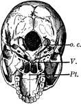
The base of the skull. The lower jaw has been removed. At the lower part of the figure is the hard palate forming the roof of the mouth and surrounded by the upper set of teeth

Base of the Skull

The base of the skull. The lower jaw has been removed. At the lower part of the figure is the hard palate…
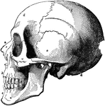
The skull. Labels: a, nasal bone; b, superior maxillary; c, inferior maxillary; d, occipital; e, temporal; f, parietal; g, frontal bone.

Human Skull

The skull. Labels: a, nasal bone; b, superior maxillary; c, inferior maxillary; d, occipital; e, temporal;…
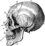
A side view of the skull. Labels: O, occipital bone; T, temporal; Pr, parietal; F, frontal; S, sphenoid; Z, malar; Mx, maxilla; N, nasal; E, ethmoid; L, lachrymal; Md, inferior maxilla.

Side View of the Skull

A side view of the skull. Labels: O, occipital bone; T, temporal; Pr, parietal; F, frontal; S, sphenoid;…