"Diagram of the eye. C., Cornea; a.h., aqueous humour; c.b., ciliary body; l., lens; I., iris; Sc., sclerotic; Ch., choroid; R., retina; v.h., vitreous humour; y.sp., yellow spot; n., optic nerve." -Thomson, 1916

Eye

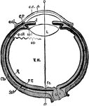
"Diagram of the eye. C., Cornea; a.h., aqueous humour; c.b., ciliary body; l., lens; I., iris; Sc.,…

Right orbit opened to show the nerves of the eye. Labels: a, optic; b, motor oculi; c, pathetic; d, ophthalmic division of fifth pair; d', lachrymal; d", supraorbital; d"', nasal; c, abducens; e', rectus externus muscle; f, orbital branch os superior maxillary nerve.

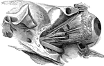
Eye Nerves of a Horse

Right orbit opened to show the nerves of the eye. Labels: a, optic; b, motor oculi; c, pathetic; d,…
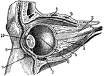
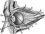
"Section through the left eye, closed. 1, lifting muscle; 2, upper straight muscle; 3, optic nerve; 4, fatty cushion; 5, lower straight muscle; 6, vitreous humor; 7, lower cross muscle; 8, lower eyelid; 9, upper eyelid; 10, crystalline lens." -Foster, 1921

Eye Section

"Section through the left eye, closed. 1, lifting muscle; 2, upper straight muscle; 3, optic nerve;…
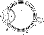
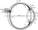
Plan of the eye seen in section. Labels: A, The Sclerotic Coat; B, The Choroid Coat; C, The Retina; D, The Optic Nerve; E, The Cornea; F, The Aqueous Humor; G, The Iris; H, The Ciliary Processes; I. The Crystalline Lens; K, The Vitreous Humor.

Diagram of the Eye

Plan of the eye seen in section. Labels: A, The Sclerotic Coat; B, The Choroid Coat; C, The Retina;…
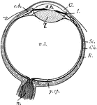
"Diagrammatic horizontal section of the eye of man. c, cornea; ch. choroid (dotted); C. P, ciliary processes; e. c, epithelium of cornea; e. cj, conjunctiva; f. c, yellow spot; I, iris; L, lens; ON, optic nerve; OS, ora serrata; o-x, optic axis; p. c. R, anterior non-visual portion of retina; P. E, pigmented epithelium (black); R, retina; sp. l, suspensory ligament; Scl, sclerotic; V. H, vitreous body." -Parker, 1900

Human Eye

"Diagrammatic horizontal section of the eye of man. c, cornea; ch. choroid (dotted); C. P, ciliary processes;…

Section through the closed left eye.1. Lifting muscle 2. Upper Straight Muscle 3. Optic Nerve 4. Fatty Cushion 5. Lower straight muscle 6. Vitrous Humour 7. Lower cross muscle 8. Lower Eyelid 9. Upper eyelid 10. Crystalline lens

Eyeball

Section through the closed left eye. 1. Lifting muscle 2. Upper Straight Muscle 3. Optic Nerve 4. Fatty…

This diagram shows a side view of the right eye of man. a.c., central artery; a.h., aqueous humor; b., blind spot, the entrance of the optic nerve; c, conjunctiva; ch., choroid layer of the eye-ball; c.l., crystalline lens; c.m.c., circular fibers of the ciliary muscles; c.m.r., radial fibers of the ciliary muscles; co., cornea, the transparent portion of the sclerotic; c.p., ciliary process; c.s., canal of Schlemm, a lymphatic vessel; fo., fovea centralis, the point of clearest vision; o.n., optic nerve; o.s., ora serrata, the anterior wavy margin of the visual portion of the retina; r, the retinal layer; sc., sclerotic layer; sh., sheath of theoptic nerve; v.h., vitreous humor.

Human Eye

This diagram shows a side view of the right eye of man. a.c., central artery; a.h., aqueous humor; b.,…
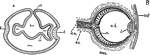
"Diagrams illustrating two stages in the development of the vertebrate eye. A, showing the relation of the ectoderm, the brain vesicle, and the optic vesicle. The right side of the figure shows a later stage than the left. B, later stage, showing the lens, eye-ball and retina in position. b.v., brain vesicle formed by the invagination oof the ectoderm (ect.); l, lens; mes., mesodermal tissue; o.n., optic nerve; o.s., optic stalk; o.v., optic vesicle, a portion of the brain vesicle; r, retinal layer; v.h., interior of eye-ball which comes to contain the vitreous humor." — Galloway

Vertebrate Eye

"Diagrams illustrating two stages in the development of the vertebrate eye. A, showing the relation…