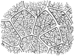
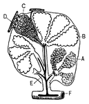
"Diagram showing the capillary network of the air sacs and origin of the pulmonary veins.. <em>A</em>, small branch of pulmonary artery; <em>B</em>, twigs of pulmonary artery; <em>C</em>, capillary network around the walls of the air sacs; <em>D</em>, branches of network converging to form the veinlets of the pulmonary veins." — Blaisedell, 1904

Capillaries of the Air Sac

"Diagram showing the capillary network of the air sacs and origin of the pulmonary veins.. A,…
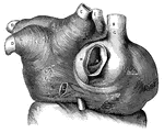
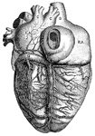
<em>L.A.</em>, left auricle; <em>R.A.</em>, right auricle; <em>A</em>, opening of the inferior vena cava; <em>B</em>, superior vena cava; <em>C</em>, cardiac vein of right auricle laid open; <em>D</em>, left pulmonary veins; <em>E</em>, eight pulmonary veins; <em>F</em>, muscular fibers surrounding the openings between the left auricle and left ventricle, and the right auricle and right ventricle; <em>G, H, K,</em> and <em>L</em>, muscular fibers surrounding the great veins of the heart." &mdash; Blaisedell, 1904

Muscular fibers of the auricle

L.A., left auricle; R.A., right auricle; A, opening of the inferior vena…
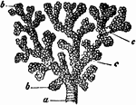
A small bronchial tube. Labels: a, dividing into its terminal branches, c; these have pouched or sacculated walls and end in the sacculated infundibula, b.

A Bronchial Tube

A small bronchial tube. Labels: a, dividing into its terminal branches, c; these have pouched or sacculated…

"<em>R.A.</em>, right auricle; <em>L.A.</em>, left auricle; <em>R.V.</em>, right ventricle; <em>L.V.</em>, left ventricle; <em>P.A.</i., pulmonary artery; <em>A</em>, pulmonary artery and vein of right lung; <em>B</em>, pulmonary artery and vein of left lung; <em>C</em>, carotid artery to head, showing branch of left subclavian artery; <em>D</em>, portal vein; <em>F</em>, hepatic artery; <em>G</em>, superior vena cava, bringing blood from head and upper limbs to right auricle." &mdash; Blaisedell, 1904

Diagram of the circulation of the blood

"R.A., right auricle; L.A., left auricle; R.V., right ventricle; L.V.,…

This illustration shows the gills (breathing apparatus) of a perch, as well as the specific veins and arteries running along the branchial arch.

Gills (Branchial Arch of Perch)

This illustration shows the gills (breathing apparatus) of a perch, as well as the specific veins and…

This illustration shows the thorax of a crayfish with a portion of the carapace removed to show the gills.

Gills (Crayfish)

This illustration shows the thorax of a crayfish with a portion of the carapace removed to show the…
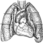
1, The trachea or windpipe; 2 and 3, right and left common carotid arteries; 4 and 5, right and left sub clavian arteries; 6, innominate artery; 7 and 8, right and left innominate veins; 9, Superior vena cava; 10, arch of the aorta; 11, ascending aorta; 12, pulmonary artery; 13, right atrium and auricula; 14 and 15, right and left lung; 16, conus arteriosus; 17, right ventricle; 18, left ventricle.

Heart and Lungs

1, The trachea or windpipe; 2 and 3, right and left common carotid arteries; 4 and 5, right and left…
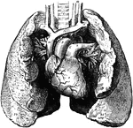
The heart and lungs. 1, right ventricle; 3, right auricle (atrium); 6, 7, pulmonary artery; 9, aorta; 10, superior vena cava; 11, innominate artery; 12, right subclavian vein; 14, innominate vein; 15, left common carotid; 17, trachea; 20, pulmonary veins; 22 to 25, lungs, partially turned back to show veins on left side.

Heart and Lungs

The heart and lungs. 1, right ventricle; 3, right auricle (atrium); 6, 7, pulmonary artery; 9, aorta;…
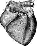
Anterior view of the heart, dissected, after long boiling to show the superficial muscular fibers. The aorta (b') and pulmonary artery (a') have been cut short close to the semilunar valves. Labels: a, right ventricle; b, left ventricle; c, c, groove between ventricles; d, d', right auricle (atrium); e, e', left auricle (atrium); f, superior vena cava; g', g", right and left pulmonary veins. The fibers are seen running a circular, oblique, transverse, and longitudinal direction.

Anterior View of the Heart

Anterior view of the heart, dissected, after long boiling to show the superficial muscular fibers. The…

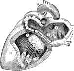
"<em>A, B</em>, right pulmonary veins, <em>S</em>, openings of the left pulmonary veins; <em>E, D, C, aortic valves; <em>R</em>, aorta, <em>P</em>pulmonary artery, <em>O</em>, pulmonic valves, <em>H</em>, mitraal valve; <em>K</em>, columnae carneae, <em>M</em> right ventricular cavity." &mdash; Blaisedell, 1904

Cavities of the heart

"A, B, right pulmonary veins, S, openings of the left pulmonary veins; E, D, C,

Left side of heart. Labels: 1, cavity of left auricle (atrium); 3, opening of right pulmonary veins; 5, left pulmonary veins; 5, left pulmonary veins; 6, auriculo-ventricular opening; 8, wall of left ventricle; 9, cavity of left ventricle; a, mitral valve (its flaps are attached by the chordae tendineae to the muscular pillars (b,b); d, arch of aorta); e, pulmonary artery.

Left Side of Heart

Left side of heart. Labels: 1, cavity of left auricle (atrium); 3, opening of right pulmonary veins;…
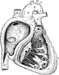
"<em>L.A.</em>, left auricle; <em>R.A.</em>, right auricle; <em>R.V.</em>, right ventricle; <em>A</em>, opening of the inferior vena cava; <em>B</em>, superior vena cava; <em>C</em>, right pulmonary veins; <em>D</em>, left pulmonary veins; <em>E</em>aorta; <em>F</em>, left branch of pulmonary artery; <em>G</em>, great cardiac vein; <em>H</em>middle cardiac vein. the cardiac lymphatics which follow the course of the cardiac veins are also shown." — Blaisedell, 1904

Posterior View of the Heart

"L.A., left auricle; R.A., right auricle; R.V., right ventricle; A,…

Right side of heart. Labels: A, cavity of right ventricle; B, superior vena cava; C, inferior vena cava; a, wall of right ventricle; b, c, columnae carneae; d, pulmonary vein; e, f, tricuspid valve; m, semilunar valve; o, wall of left ventricle; p, q, v, ascending aorta, arch and descending aorta.

Right Side of Heart

Right side of heart. Labels: A, cavity of right ventricle; B, superior vena cava; C, inferior vena cava;…
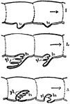
"Diagram to show the way in which an outgrowing gill-process bearing blood-holding lamellae, may give rise, if the sternal body wall sings inwards, to a lung-chamber with air-holding lamellae. I is the embryonic condition. L is the condition of outgrowth with gl, gill lamellae. A is the condition of insinging of the sternal surface and consequent enclosure of the lamelligerous surface of the appendage in a chamber with narrow orifice&mdash;the pulmonary air-holding chamber. pl, Pulmonary lamellae. bs, Blood sinus." &mdash; The Encyclopedia Britannica, 1910

Lamellae

"Diagram to show the way in which an outgrowing gill-process bearing blood-holding lamellae, may give…

"Section of a pulmonary lobule, showing its division into pulmonary vesicles." &mdash; Tracy, 1888

Pulmonary lobule

"Section of a pulmonary lobule, showing its division into pulmonary vesicles." — Tracy, 1888

"<em>A</em>, an air sac; <em>B</em>, an air sac cut open; <em>C</em>, capillary network over an air sac; <em>D</em>, branch of pulmonary artery; <em>E</em>, branch of pulmonary vein; <em>F</em>, bronchial tube." &mdash; Blaisedell, 1904

Lobule of the lung

"A, an air sac; B, an air sac cut open; C, capillary network over an air…

The lungs and air passages seen from the front. On the left of the figure the pulmonary tissue has been dissected away to show the ramifications of the bronchial tubes. Labels: a, larynx; b, trachea; d, right bronchus. The left bronchus is seen entering the root of its lung.

Lungs and Air Passages

The lungs and air passages seen from the front. On the left of the figure the pulmonary tissue has been…

The lungs and air passages seen from the front. On the left of the figure the pulmonary tissue has been dissected away to show the ramifications of the bronchial tubes. Labels: a, larynx; b, trachea; d, right bronchus. The left bronchus is seen entering the root of its lung.

The Lungs and Air Passages

The lungs and air passages seen from the front. On the left of the figure the pulmonary tissue has been…
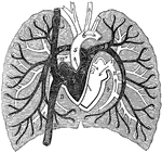
A diagram of pulmonary circulation. Labels: 1, Descending vena cava. 2, Ascending cava vein. 3, Chamber of right side of heart. 4, Chamber of left side of heart. 5, Aorta. 6, Arch of aorta. 7, Pulmonary artery. 8, Its left branch. 9, Its right branch. 10, Right pulmonary vein. 11, Left pulmonary vein.

A Diagram of Pulmonary Circulation

A diagram of pulmonary circulation. Labels: 1, Descending vena cava. 2, Ascending cava vein. 3, Chamber…
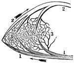
A portion of the pulmonic circulation. 1, A branch of the artery that carries the impure blood to the lungs. 3, Capillary vessels. 2, A vein through which red blood is returned to the left side of the heart.

A Portion of the Pulmonic Circulation

A portion of the pulmonic circulation. 1, A branch of the artery that carries the impure blood to the…

"Embryo of scorpion, ventral view showing somites and appendages. sgc, Frontal groove. sa, Rudiment of lateral eyes. obl, Camerostome (upper lip). so, Sense-organ of Patten. PrGabp1, Rudiment of the appendage of the praegenital somite which disappears. abp2, Rudiment of the right half of the genital operculum. abp3, Rudiment of the right half of the genital operculum. abp4 to abp7, Rudiments of the four appendages which carry the pulmonary lamellae. I to VI, Rudiments of the six limbs of the prosoma. VIIPrG, The evanescent praegenital somite. VIII, The first mesosomatic somite or genital somite. IX, The second mesosomatic somite or pectiniferous somite. X to XIII, The four pulmoniferous somites. XIV, The first metasomatic somite." &mdash; The Encyclopedia Britannica, 1910

Scorpio Embryo

"Embryo of scorpion, ventral view showing somites and appendages. sgc, Frontal groove. sa, Rudiment…
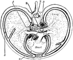
"Cross-section of thorax. A, bronchus, entering the lung; B, the aorta cut at its origin and again at the descending part of its arch; C, the pericardial space; D, the pleural cavity; E, the alimentary canal; PA, the pulmonary artery; X, the neural canal." &mdash;Martin, 1917

Thorax

"Cross-section of thorax. A, bronchus, entering the lung; B, the aorta cut at its origin and again at…
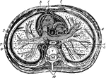
Transverse section of the thorax. Labels: 1, anterior mediastinum; 2, internal mammary vessels; 3, triangularis sterni; 4, 5, phrenic nerves; 6, thoracic duct; 7, esophagus; 8, vena azygos major; 9, thoracic aorta; 10, sympathetic; R. V., right ventricle; R. A., right auricle; P. A. pulmonary artery; A., aorta; C., superior vena cava; V., dorsal vertebra.

Transverse Section of the Thorax

Transverse section of the thorax. Labels: 1, anterior mediastinum; 2, internal mammary vessels; 3, triangularis…