Sphenoid bone, situated the anterior part of the base of the skull, articulating with all the other cranial bones, which it binds firmly and solidly together. Labels: a, greater wing; b, lesser wing.

Sphenoid Bone of the Human Skull

Sphenoid bone, situated the anterior part of the base of the skull, articulating with all the other…
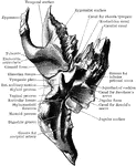
The sphenoid as seen from behind.

Sphenoid Viewed from Behind

The sphenoid as seen from behind.

The sphenoid as seen from the front.

Sphenoid Viewed from the Front

The sphenoid as seen from the front.

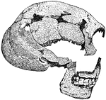
"One of two skulls discovered in 1886 in the cave of Spy (Belgium). Notice the prominent eyebrow ridges, the low, retreating forehead, the strong and well-developed lower jaw."—Webster, 1913

Skull of the Man of Spy

"One of two skulls discovered in 1886 in the cave of Spy (Belgium). Notice the prominent eyebrow ridges,…
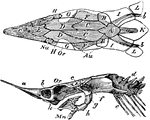
"Top and side views. The cartilaginous cranium shaded, is supposed to be seen through the unshded canial bones."-Whitney, 1902

Sturgeon Skull

"Top and side views. The cartilaginous cranium shaded, is supposed to be seen through the unshded canial…
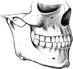
To show the relation of the upper to the lower teeth when the mouth is closed. The manner in which a tooth of one row usually strikes against two teeth of the opposite row, and the resulting interlocking of the teeth, is to be noted.

Teeth

To show the relation of the upper to the lower teeth when the mouth is closed. The manner in which a…
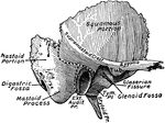
The outer surface of the temporal bone. The dotted lines indicate the lines of suture between squamous, mastoid, or tympanic portions.

Temporal Bone

The outer surface of the temporal bone. The dotted lines indicate the lines of suture between squamous,…

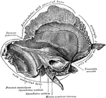
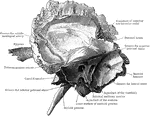
The inner surface of the left temporal bone at birth.

Temporal Bone at Birth

The inner surface of the left temporal bone at birth.

A, The outer surface of the right temporal bone at birth. B, The same with squamozygomatic portion removed. Labels: a, tympanic ring; b, inner wall of tympanum; c, fenestra rotunda; d, foramen ovale; e, mastoid; f, mastoid process; g, mastosquamosal suture, with foramen for transmission of vessels; h, squamozygomatic, removed in figure B to show how its descending process forms the outer wall of the mastoid antrum.

Temporal Bone at Birth

A, The outer surface of the right temporal bone at birth. B, The same with squamozygomatic portion removed.…
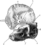
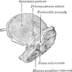
Inner surface of right temporal bone at birth. am squamozygomatic; b, petrosquamosal suture and foramen (just above the end of the lead line; c, subarcuate fossa; d, aquaedutcus vestibuli; e, aquaeductus cochlea; f, internal auditory meatus; g, upper end of carotid canal.

Temporal Bone at Birth

Inner surface of right temporal bone at birth. am squamozygomatic; b, petrosquamosal suture and foramen…
Temporal bone at birth. Inner aspect.

Inner Aspect of Temporal Bone at Birth

Temporal bone at birth. Inner aspect.

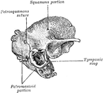
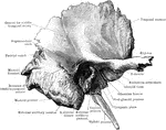
Temporal bone at birth. Outer aspect.

Outer Aspect of Temporal Bone at Birth

Temporal bone at birth. Outer aspect.

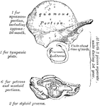
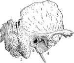
Temporal bone of the human skull. The temporal bones are situated at the sides and base of the skull. Labels: 1, squamous portion; 2, placed below external opening of auditory canal in petrous portion; 3, placed below mastoid portion; 4, placed below glenoid cavity for reception of condyle of lower jaw.

Temporal Bone of the Human Skull

Temporal bone of the human skull. The temporal bones are situated at the sides and base of the skull.…
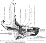
The right temporal bone as seen from below.

Temporal Bone Viewed from Below

The right temporal bone as seen from below.

The anterior half of a vertical transverse section through the left temporal bone.

Anterior Half of Section Through Temporal Bone

The anterior half of a vertical transverse section through the left temporal bone.
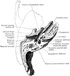
Development of the temporal bone. From ten centers.

Development of Temporal Bone

Development of the temporal bone. From ten centers.

Horizontal section through left temporal bone showing lower half of section.

Horizontal Section of Temporal Bone

Horizontal section through left temporal bone showing lower half of section.
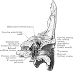
The right temporal bone as seen from the inner side.

Inner Surface of the Temporal Bone

The right temporal bone as seen from the inner side.

The right temporal bone as seen from the outer side.

Outer Surface of the Temporal Bone

The right temporal bone as seen from the outer side.

The posterior half of a vertical transverse section through the left temporal bone.

Posterior Half of Section Through Temporal Bone

The posterior half of a vertical transverse section through the left temporal bone.

"Skull of Tillotherium Fodiens." —The Encyclopedia Britannica, 1903

Tillotherium Fodiens

"Skull of Tillotherium Fodiens." —The Encyclopedia Britannica, 1903

An illustration of a woodpecker's skull.

Woodpecker Skull

An illustration of a woodpecker's skull.