Clipart tagged: ‘spinal’
Brain
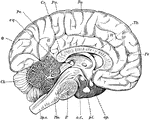
"Diagram of the left half of a vertical median section of the brain. H, H, convoluted inner surface…

Dissected Fish
"Dissected fish. a, air bladder; b, urinary bladder; b, urinary bladder; br, brain; c, spinal cord;…

Dissected Frog
"Frog with the left side cut away and some of the organs pulled downward. a, aorta leading from the…
Medulla Oblongata
"The spinal cord and medulla oblongata. A, from the ventral, and B, from the dorsal aspect; C to H,…
Nerve Roots
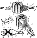
"The spinal cord and nerve-roots. A, a small portion of the cord seen from the ventral side; B, the…

Perch Skeleton
"The spinal column consists of abdominal and caudal vertebre, the coalescence of the parapophyses into…
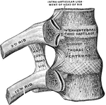
Ribs and Corresponding Vertebral Bodies
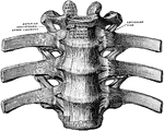
Ribs and corresponding vertebral bodies in their ligaments, viewed from the right.
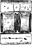
Shark Vertebra
"Lateral view of caudal vertebra of Basking Shark (Selache mazima). a, centrum; b, neurapophysis; c,…
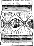
Shark Vertebra
"Longitudinal section of caudal vertebra of Basking Shark (Selache mazima). a, centrum; b, neurapophysis;…

Shark Vertebra
"Transverse section of caudal vertebra of Basking Shark (Selache mazima). a, centrum; b, neurapophysis;…

Spinal Accessory Nerve
Scheme of the origin, connection, and distribution of the spinal accessory nerve. Labels: Sp.Acc, spinal…
Spinal Column
The spinal column. 1, 2, and 3: Vertebrae. 4 and 5: The sacrum and cocyx bones of the pelvis. 6: Processes.
Spinal Column
Side view of the spinal column, with the vertebrae numbered: C1-7, cervical vertebrae; D1-12, dorsal…
The Spinal Column
View of the entire spinal column. The bodies of the vertebrae are in the front with the spinous processes…

Section of Spinal Cord
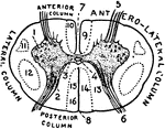
"Diagram of a slice across the spinal cord, showing the roots of a spinal nerve to the arm on the left.…
Spinal Divisions
A lateral view of the spine divided into its cervical, dorsal, and lumbar portions.

Spine
Diagram on frozen section, showing relations of bodies and spines of vertebrae to levels at which spinal…
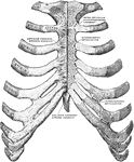
Sternum and Ribs
Sternum and ribs with ligaments, from in front. In the right half of the figure the most anterior layer…

Lateral and Dorsal View of the Vertebral Column
The spinal column, right lateral view and dorsal view.

