Search for "retinal"
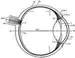
Human Eye
This diagram shows a side view of the right eye of man. a.c., central artery; a.h., aqueous humor; b.,…
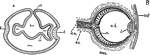
Vertebrate Eye
"Diagrams illustrating two stages in the development of the vertebrate eye. A, showing the relation…
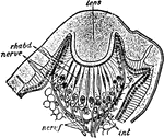
Euscorpius Italicus
"Section through the lateral eye of Euscorpius italicus. lens, Cuticular lens. nerv.c, Retinal cells…
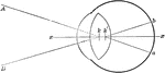
Retina
"Diagram illustrating the points at which incident rays meet the retina. xx, optic axis; k, first nodal…
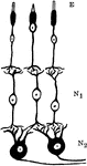
Neurons and Sensory Epithelium in the Retina
Diagram showing relations of the neurons and sensory epithelium in the retina. labels: E, epithelial…

Retina Blind Spot
The right retina as it would be seen if the front part of the eyeball with the lens and vitreous humor…
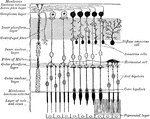
Retinal Structure
Diagram of the structure of the human retina. Labels: I, pigment layer; II, rod and cone layer; R, rods;…
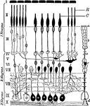
Invertebrate Simple Eye
"Diagrams showing some of the stages in the increasing complexity of the simple eye in Invertebrates.…
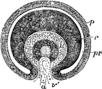
Eye of Fetus of Four Weeks
Transverse vertical section of the eyeball of a human embryo of four weeks. The anterior half of the…
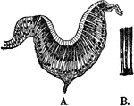
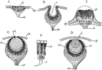
Eyepit of Limpet
A, section through the open eyepit of a limpet. B, the two kinds of retinal cells, pigmented and sensory.