Development of the Alimentary Canal
To use any of the clipart images above (including the thumbnail image in the top left corner), just click and drag the picture to your desktop. You may also control-click (Mac) or right-click (Windows) and choose "Save file to disk" from the pop-up menu. Click here for help downloading and using clipart files.
* A TIFF file is a high-resolution (240 dpi) format. TIFF files should be used only if you are printing the clipart. Do NOT use TIFF files for clipart that will displayed on a computer screen. Click here for help downloading and using TIFF files.File Name: 51318_alimentary
Description: 
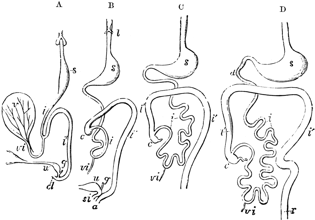
Outlines of the form and position of the alimentary canal in successive stages of its development. A, alimentary canal, in an embryo of four weeks; B, at six weeks; C, at eight weeks; D, at ten weeks; l, the primitive lungs connected with the pharynx; s, the stomach; d, duodenum; i, the small intestine; i', the large intestine; c, the caecum and vermiform appendage; r, the rectum; cl, in A, the cloaca; a, in B, the anus distinct from s i, the sinus uro-genitalis; v, the yolk sac; vi, the vitellointestinal duct; u, the urinary bladder and urachus leading to the allantois; g, genital ducts.
Source: Baker, W. Morrant & Harris, Vincent Dormer Kirkes' Hand-book of Physiology, 13th ed. (Philadelphia: P. Blakiston's Son & Co., 1892) 840
Keywords: "alimentary canal", digestive, digestion
Copyright: 2009, Florida Center for Instructional Technology. See license.
Main Menu Site Map Search License Clipart Help
Clipart ETC is a part of the Educational Technology Clearinghouse and is funded by various grants. Produced by the Florida Center for Instructional Technology, College of Education, University of South Florida. Email the project manager.