Clipart tagged: ‘digestion’
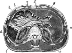
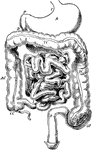
Horizontal Section Through Abdomen
Horizontal section through upper part of abdomen. Labels: a, liver; b, stomach; c, transverse colon;…
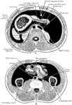
Transverse Section of Abdomen
Diagrammatic transverse section of abdomen, to show the peritoneum on transverse tracing. A, at level…
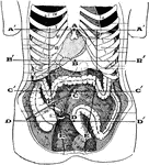
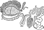
Abdominal Region
Showing the average position of the abdominal viscera with their surface markings. Labels: A, sterno-ensiform…
Alimentary Canal
Diagram of the abdominal part of the alimentary canal (digestive system). Labels: C, the cardiac, and…
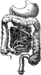
Alimentary Canal
Diagram of the abdominal part of the alimentary canal. Labels: C, the cardiac, and P, the pyloric end…
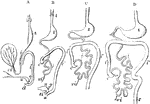
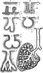
Development of the Alimentary Canal
Outlines of the form and position of the alimentary canal in successive stages of its development. A,…
Development of the Alimentary Canal
Front view of two successive stages in the development of the alimentary canal.
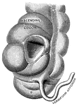
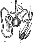
Vermiform appendix
"A, a portion of the colon laid open to show the valve between the large and small intestine;…
The Digestive Apparatus of a Beetle
The Annulosa and Mollusca are furnished with a distinct alimentary canal that does not open into the…
Blood Circulation
This illustration shows a representation of the circulation of the blood, in its essential features.…
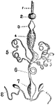
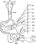
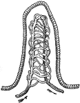
Intestinal Tract from Canis Vulpes
Intestinal tract of Canis vulpes. S, cut end of duodenum; C, caecum; R, cut end of rectum.
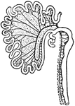
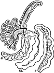
Intestinal Tract of Chauna Chavaria
cc. Colic caeca, d. Duodenum. g. Glandular patch, l.l. Meckel's tract, l.i. Hind-gut, p.v. Cut root…
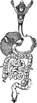
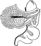
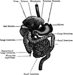
Digestive Organs
The stomach, pancreas, liver, and duodenum, with part of the rest of the small intestine and the mesentery;…
Digestive System
A diagram of the organs of digestion. Labels:1, The upper jaw. 2, The lower jaw. 3, The tongue. 4, The…
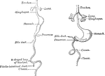
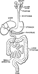
Digestive Tract
This is a diagram of the digestive tract. Notice how the food is submitted to the action of alkaline,…

Digestive Tract of a Chick
Diagram of part of digestive tract of a chick (4th day). The black line represents hypoblast , the outer…
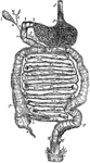
Fowl Digestive System
"Digestive system of the common Fowl. o, Gullet; c, Crop; p, Proventriculus; g, Gizzard; sm, Small intestine;…
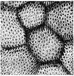
Gastric gland
"The inner coat of the stomach has its surface honeycombed with millions of little pits. We have all…
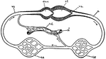
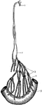
Intestinal Tract of Giraffe
S, cut end of duodenum; R, cut end of rectum; C, caecum; P.C.L., post-caecal loop; S.P., spiral loop;…
Forms of Glands
Forms of glands. Labels: A, a simple secreting surface; a, its epithelium; b, basement membrane; c,…
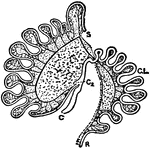
The Digestive Apparatus of a Hydra or Freshwater Polyp
In Radiata the digestive cavity is a pouch with a single opening, into which the food is passed and…
Intestinal absorption
"A, a fold of peritoneum; B, lacteals and lymphatic glands; C, veins of intestines;…
Intestinal Tract of Macropus Bennetti
S, cut end of duodenum; R, cut end of rectum; C, caecum; C2, accessory caecum; C.L., colic loop of hind-gut.
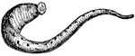
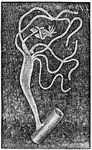
Medicinal Leech
The medicinal leech is a leech used in bloodletting. It lives in fresh water, and is common in Germany,…
Nerves of the Alimentary Canal
Diagrammatic representation of the nerves of the alimentary canal. Oe to Rct, the various parts of the…
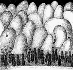
Glands and villi of the small intestine
"A, B, glands seen in vertical section with their orifices at C opening upon the membrane…
The Small Intestine
The small intestine, a convoluted, tubular, digestive organ, about 20 ft in length, extending from the…
Transverse section of the small intestine
"In the figure on the left are seen the artery and vein of a villus. In the right figure are represented…
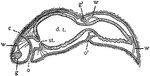
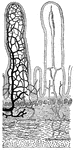
Stenostoma
In this Turbellarian the digestive tract (d.t.) is a blind sac. st., boundary of stomodaeum and mesenteron;…

Stomach
"The stomach is a half-gallon sac, with an outer wall of muscle lined within by mucous membrane, made…
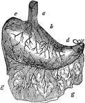
Stomach
The human stomach. Labels: a, the esophagus or gullet; b, the cardiac portion; c, the left extremity;…
Stomach
The digestive system. This figure represents the whole tract of the intestinal canal, not exactly in…
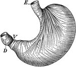
Stomach
The stomach showing the muscles which churn the food. Labels: E, where food enters; V, entrance into…
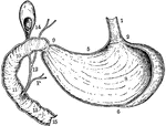
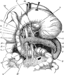
Vertical and Longitudinal Section of Stomach, Gall-Bladder, and Duodenum
Vertical and longitudinal section of stomach, gall-bladder, and duodenum. Labels: 1, esophagus; 2, cardiac…
Inner surface of the stomach
"The Inner Surface of the Stomach, from which the the Epithelium has been removed, showing the Openings…
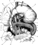
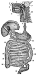
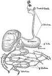
The Stomach, Pancreas, Liver, and Duodenum
The stomach, pancreas, liver, and duodenum, with part of the rest of the small intestine and the mesentery;…
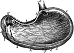
The Stomach
The stomach, the principal organ of digestion. It is a dilated part of the alimentary canal, situated…
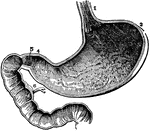
The Stomach
The inside of the stomach with the beginning of the intestines. At 3 is the left end and at 4 is the…
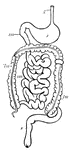
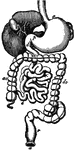
Digestive system
Digestive system of a mammal. (g) gullet; (s) stomach; (sm) small intestine; (lm) large intestine; (r)…
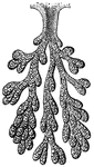
Thoracic duct and lacteals
"The lacteals conduct the chyle from the intestines into numerous glands nearby, called the mesenteries,…
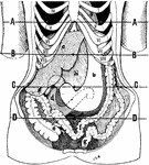
Position of the Viscera in the Condition of Visceroptosis
Showing the position of the viscera in the condition of visceroptosis (Glenard's disease). Labels: A,…