This human anatomy ClipArt gallery offers 82 illustrations of human teeth and jaws. This includes external and internal views focusing on human adult and juvenile teeth. Views of teeth within the jaw bones are also included.
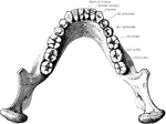
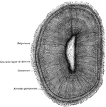
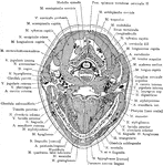
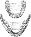
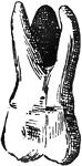
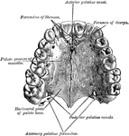
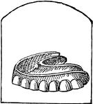
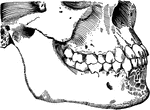
Back view of the adult mouth
"The head is represented as having been thrown back, and the tongue drawn forward. A, B…
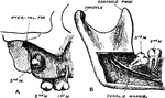
Antrum of Highmore with Roots of Teeth
The alveolar process and teeth were ground off until the antrum of highmore was well exposed; its anterior…
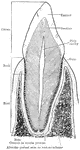
Structure of Canine Tooth
Vertical section of canine tooth to illustrate the various parts and structures.
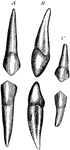
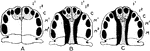
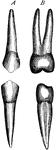
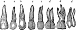
Temporary Canine
Canine teeth of left side, labiial (A) and lateral (B) aspects. C, temporary canines.
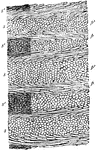
Dentine and Cement
Section of a portion of the dentine and cement from the middle of the root of an incisor tooth. Labels:…
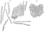
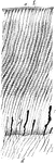
Enamel Fibers
Enamel fibers. A, fragments and single fibers of the transversely striated enamel, isolated by the action…
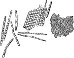
Enamel Prisms
Enamel prisms. A, Fragments and single fibers of the enamel isolated by the action of hydrochloric acid;…
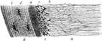
Longitudinal Section of Enamel
Longitudinal section of enamel, treated with acid, showing disposition of ranges of enamel prisms (p,…
Section of the Enamel
Enamel is composed of fine hexagonal fibers which are set on end on the surface of the dentine and fit…
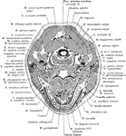
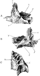
Cross Section of Head Through the Body of the Mandible
Section of the head through the body of the mandible.
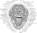
Cross Section of Head Through the Inferior Portion of the Mandible
Section of the head through the inferior portion of the mandible.
Incisor Relation to Palatal Cleft
Illustrating the relationship of the lateral incisor tooth to the palatal cleft. A, Normal hard palate.…
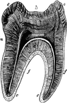
Jaw Showing Roots of Teeth
Horizontal section through both the upper and lower jaws to show the roots of the teeth. The sections…
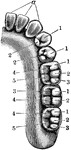
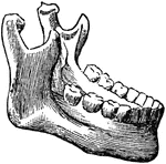
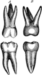
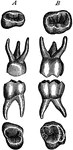
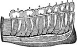
Lower Jaw with Teeth
Right half of lower jaw, with the corresponding teeth. The letters and numbers point to the various…
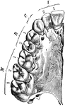
Upper Jaw with Teeth
Right half of upper jaw (from below), with the corresponding teeth. The letters and numbers point to…
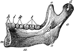
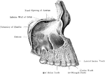
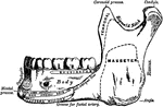
Lower Jaw
Half of the lower jaw. Labels: a, the base; b, the angle; c, the ramus; d, the condyle; e, the coronaid…

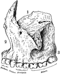
Ossification of Superior Maxilla
Ossification of superior maxilla. A, outer side. B, inner side. C, under side. Labels: a, nasal process;…
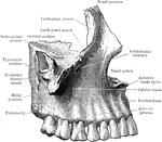
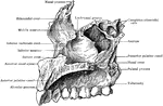
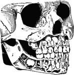
Human Maxillary (Upper Jaw) Bone
Superior maxillary bone. With it's fellow on the opposite side, it forms the whole of the upper jaw.…
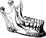
Human Maxillary (Upper Jaw) Bone
Inferior Maxillary Bone (lower jaw). It is the largest and strongest bone in the face and serves for…
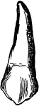
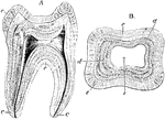
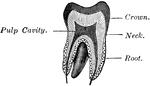
Molar Tooth
Longitudinal section of a molar tooth. Labels: k, crown; n, neck; f, fangs; e, enamel; d, dentine; c,…
Impacted Third Molar
Impaction of the upper third molar in the maxilla. B, Impaction of the lower third molar in the mandible.
Structure of a Molar
Longitudinal section (A) and transverse section (B) of a human molar tooth. Labels: c, cement; d, dentine;…
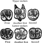
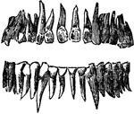
Surface of Molar
Triturating surfaces of molar teeth of right side. The upper margin of the figures corresponds to the…
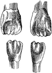
Temporary Molar
Temporary molar teeth (A, first; B, second) of left side. Triturating surfaces of crowns also shown.
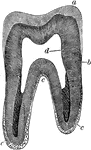
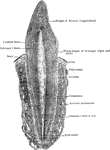
Vertical Section of a Molar
Vertical section of a molar tooth. Labels: a, enamel of the crown, the line of which indicate the arrangement…
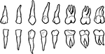
Teeth
The series of small bones attached to the jaws of animals, or human beings, which serve the purpose…
Teeth
Image of teeth in a human jaw. "1, incisors; 2, canine; 3, bicuspids; 4, molars (the molar at the left…
Teeth
To show the relation of the upper to the lower teeth when the mouth is closed. The manner in which a…
Teeth of a Child
Temporary teeth in a child aged about 4 years. The permanent teeth are seen in the process of formation…
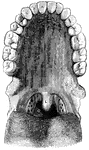
Teeth of the Upper Jaw
Half of the teeth of the upper jaw. a,a, are the two front cutting teeth. d,d,d are…
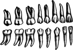
The Adult Teeth
The adult teeth. Labels: 1, 2, The cutting teeth (incisors). 3, Eyetooth (cuspid). 4,5, Small grinders…
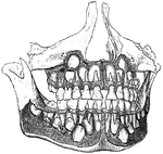
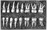
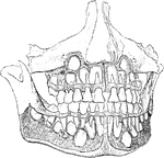
The Deciduous and Permanent Teeth
The deciduous and permanent teeth, shown as they are placed in the jaw with portions of bone removed…
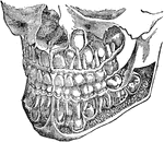
Development of Permanent Teeth
Well formed jaws, from which the alveolar plate has been removed to expose the developing permanent…
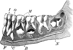
Emergence of Adult Teeth
The teeth of a 6 and a half year old child. Label: I, the incisors; O, the canine; M, the molars; the…
!["A Tooth is one of the hard bodies of the mouth, attached to the skeleton, but not forming part of it and developed from the dermis or true skin. True teeth consist of one, two, or more tissues differing in their chemical composition and in their microscopical appearances. Dentine, which forms the body of the tooth, and 'cement,' which forms its outer crust, are always present, the third tissue, the 'enamel,' when present, being situated between the dentine and cement. The incisors, or cutting teeth, are situated in front. In men there are two of these incisors in each side of each jaw. The permanent incisors, molars, and premolars are preceded by a set of deciduous or milk teeth, which are lost before maturity, and replaced by the permanent ones. The canines come next to the incisors. In man there is one canine tooth in each half-jaw. The premolars (known also as bicuspids and false molars) come next in order to the canines. In man there are two premolars in each half-jaw. The true molars (or multicuspids) are placed most posteriorly. In man there are three molars in each half-jaw, the posterior one being termed the wisdom tooth. The figures [in the illustration] refer to months after birth."—(Charles Leonard-Stuart, 1911)](https://etc.usf.edu/clipart/15200/15256/teeth1_15256_mth.gif)
First Teeth
"A Tooth is one of the hard bodies of the mouth, attached to the skeleton, but not forming part of it…