Human Smooth and Cardiac Muscle
The Smooth and Cardiac Muscle ClipArt gallery offers 26 illustrations of heart, blood vessel, gastrointestinal tract, respiratory tract, and other involuntary system muscles.
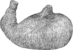
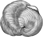
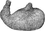
Muscular fibers of the auricle
L.A., left auricle; R.A., right auricle; A, opening of the inferior vena…

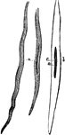
Spindle Cell of Involuntary Muscle
"The involuntary muscles consist of ribbon-shaped bands which surround hollow tubes or cavities…
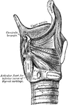
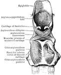
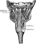
Side View of the Muscles of the Larynx
Muscles of the larynx. Side view. Right ala of thyroid cartilage removed.
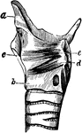
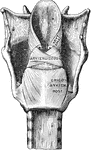
Larynx Muscle
Dissection of the muscles in the lateral wall of the larynx. The right ala of the thyroid cartilage…
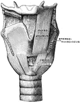
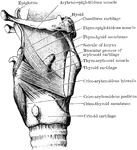
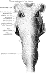
Front View of the Muscles of the Larynx
Muscles of the larynx, front view. The Sternothyroid and right Thyrohyoid have been removed.
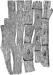
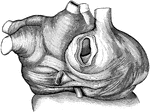
Muscle Fibers of the Heart
Anastomosing muscle fibers of the heart, seen in longitudinal section. On the right the limits of the…
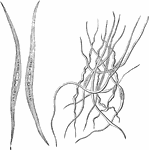
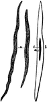
Smooth Muscle Fibers
The smooth muscle fibers consist of long spindle-shaped cells, the end of which are frequently spirally…
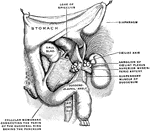
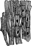
Muscle Lining of Stomach
The muscular coat (lining) of the stomach, a type of involuntary muscle involved in the contraction…
Heart Muscle
The muscular fibres of the heart, unlike those of most involuntary muscles, are striated.
Smooth Muscle
Three sections of smooth muscle. M C is muscle cells and N is nucleus. Smooth muscles are slow to contract.…
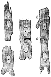
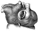
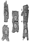
Muscular Fiber Cells from the Heart
Muscular fiber cells from the heart. The fibers which lie side by side are united at frequent intervals…
Cardiac Muscular Tissue
Cardiac muscular tissue, magnified about 400 diameters. The cell-boundaries and cell-nuclei are indicated…
Non-Striated Muscle
"Non-striped spindle-shaped cells which branch and join with one another." — Richardson, 1906
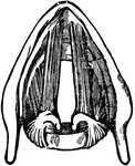
Muscles of the Pharynx
The muscles of the pharynx. On the right side most of the inferior constrictor has been removed, on…